Validation Data Gallery

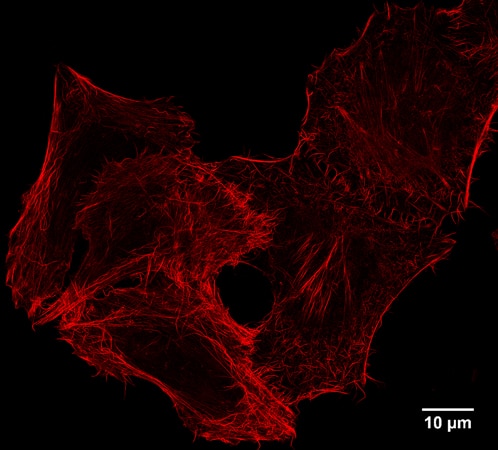
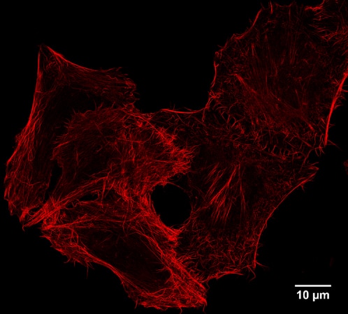
  • STED: IF of Spot-tagged Actin-Chromobody with Spot-Label Atto594 bivalent (1:1,000). Gated STED images were acquired with a Leica TCS SP8 STED 3X microscope with pulsed White Light Laser excitation at 590 nm and pulsed depletion with a 775 nm laser. Objective: 100x Oil STED White, NA: 1.4. Pixel size: 21 x 21 nm; z-Step size of z-Stacks: 0.16 μm. Images were deconvolved with Huygens Professional (SVI). STED images were recorded at the Core Facility Bioimaging at the Biomedical Center, LMU Munich.

    STED: IF of Spot-tagged Actin-Chromobody with Spot-Label Atto594 bivalent (1:1,000). Gated STED images were acquired with a Leica TCS SP8 STED 3X microscope with pulsed White Light Laser excitation at 590 nm and pulsed depletion with a 775 nm laser. Objective: 100x Oil STED White, NA: 1.4. Pixel size: 21 x 21 nm; z-Step size of z-Stacks: 0.16 μm. Images were deconvolved with Huygens Professional (SVI). STED images were recorded at the Core Facility Bioimaging at the Biomedical Center, LMU Munich.

  • Application of Spot-tag related products.

    Application of Spot-tag related products.

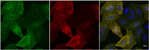
  • Widefield images of HeLa cells expressing Tom70-EGFP-Spot-Tag immunostained with Spot-Label ATTO594, bivalent (1:5,000). Left: GFP channel, middle: channel for ATTO594, right: overlay, also with DAPI. Scale bar, 10 μm.

    Widefield images of HeLa cells expressing Tom70-EGFP-Spot-Tag immunostained with Spot-Label ATTO594, bivalent (1:5,000). Left: GFP channel, middle: channel for ATTO594, right: overlay, also with DAPI. Scale bar, 10 μm.

  • Confocal images of HeLa cells expressing Tom70-EGFP-Spot-Tag immunostained with Spot-Label ATTO594 bivalent (1:1,000). Left: EGFP fluorescence, middle: ATTO594 signal, right: merge. Scale bar, 10 μm. The images were acquired with Leica TCS SP8 microscope, 100X oil objective, at the Core Facility Bioimaging at the Biomedical Center, LMU Munich, and deconvolved with Huygens Professional (SVI). Left: GFP fluorescence, middle: ATTO594 signal, right: merge. Scale bar, 10 μm.

    Confocal images of HeLa cells expressing Tom70-EGFP-Spot-Tag immunostained with Spot-Label ATTO594 bivalent (1:1,000). Left: EGFP fluorescence, middle: ATTO594 signal, right: merge. Scale bar, 10 μm. The images were acquired with Leica TCS SP8 microscope, 100X oil objective, at the Core Facility Bioimaging at the Biomedical Center, LMU Munich, and deconvolved with Huygens Professional (SVI). Left: GFP fluorescence, middle: ATTO594 signal, right: merge. Scale bar, 10 μm.

ChromoTek Spot-Label® ATTO594

anti-Spot-Tag® VHH/ Nanobody conjugated to fluorescent dye for immunofluorescence, microscopy, and immunoblotting of Spot-tagged proteins
Cat No. eba594

Specificity

Spot-Tag®

Applications

IF

Conjugate

ATTO 594

Host

Alpaca

Spot tag, Spot, PDRVRAVSHWSS, Spot-Label

Size/Concentration: 

-/ -


ご購入について

国内販売は「コスモ・バイオ株式会社」を通じて行っております。お見積り・ご注文はお近くの販売代理店へご連絡ください。


国内在庫・納期について

約2万点のプロテインテック製品をコスモバイオ社物流センター(国内)に在庫しています。国内在庫の有無はコスモバイオ社ホームページの「品番検索」でカタログ番号を検索して確認できます。


保証・サポートについて

テクニカルサポートまたはご購入後1年間の交換/補填対応を承ります。詳細はこちらをご覧ください。


Product Information

anti-Spot-Tag® VHH/ Nanobody conjugated to fluorescent dye for immunofluorescence, microscopy, and immunoblotting of Spot-tagged proteins

DescriptionImmunofluorescence of Spot-tagged proteins with anti-Spot Nanobody conjugated to fluorescent dye. Spot-Label is much smaller compared to conventional primary plus secondary IgG antibodies (IgG complex). Due to its small size, Spot-Label is optimal for effective labeling with minimal fluorophore displacement for super-resolution microscopy.

• Capture and detection tag without compromises in applications

• Higher image resolution

• Superior accessibility and labelling of epitopes in crowded cellular/organelle environments

• Strong avidity effect from bivalent form of Spot-VHH

• Fulfills highest requirements on antibody validation: structure and function characterized

ApplicationsIF
HostAlpaca
Specificity/TargetSpot-Tag® (PDRVRAVSHWSS)
ConjugateATTO 594
Physical StateLiquid
Suggested DilutionIF: 1:1,000
Type Nanobody
ClassRecombinant
RRIDAB_2827570
Affinity (KD) Dissociation constant KD of 6 nM
Spot-Tag® OriginEngineered 12 amino acid sequence modified from the unstructured N-terminus of beta-catenin
Cross ReactivityThe Spot-Nanobody shows minimal cross reactivity with beta-catenin.
Excitation/ EmissionExcitation range 580 - 615 nm (λabs= 601 nm)
Emission range 620 - 660 nm (λfl= 627 nm)
Storage Buffer1x PBS, 0.09% sodium azide
Storage ConditionShipped at ambient temperature. Upon receipt store at +4°C. Stable for 6 month. Do not freeze. Protect from light.
Size10 μL; 50 μL
NoteThis product is for research use only, not for diagnostic or therapeutic use

Publications

ApplicationTitle
IF

Microbiol Spectr

BopN is a Gatekeeper of the Bordetella Type III Secretion System

Authors - Kevin Munoz Navarrete
IF

Methods Mol Biol

Superresolution Expansion Microscopy in Dictyostelium Amoebae

Authors - Marianne Grafe
IF

Cell Death Differ

Inhibition of BAK-mediated apoptosis by the BH3-only protein BNIP5

Authors - Sebastian Rühl