Validation Data Gallery

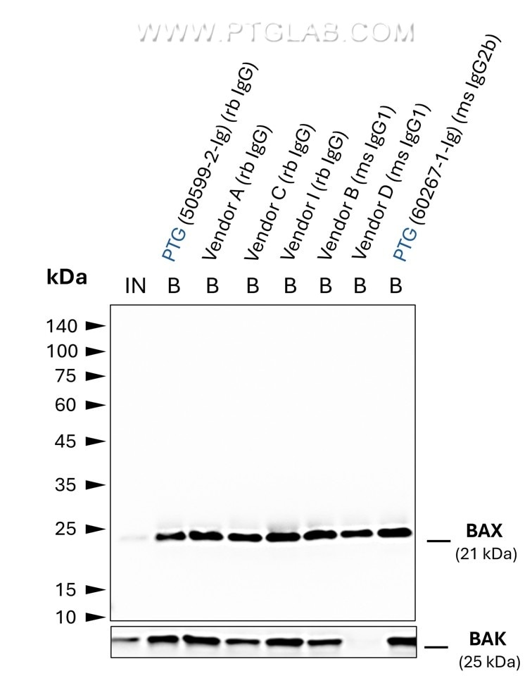
  • Co-IP of BAX and BAK using anti-BAX IgGs from different vendors by anti-rabbit IgG/ anti-mouse IgG VHH Agarose (mrIGa). Tested IgGs include such of rabbit and mouse origin with IgG1 and IgG2b subtypes. 5 µg of respective IgG was spiked into HEK293T cell lysate derived from 0.5x10^7 cells. 1% of input (IN) and 25% of bound (B) fraction was loaded onto an SDS-PAGE gel. For Western Blot analysis BAX was detected using a polyclonal rabbit IgG (PTG: 50599-2-Ig) (1:2000) labeled using a conformation-specific HRP-conjugated secondary. The presence of BAK co-precipitated with BAX was tested using a polyclonal rabbit IgG (PTG 29552-1-AP) (1:2000). While all IgGs allowed for efficient IP of BAX, BAK was absent in the case of the primary antibody from vendor D, possibly due to the BAX:BAK interaction interface being masked by this particular IgG.

    Co-IP of BAX and BAK using anti-BAX IgGs from different vendors by anti-rabbit IgG/ anti-mouse IgG VHH Agarose (mrIGa). Tested IgGs include such of rabbit and mouse origin with IgG1 and IgG2b subtypes. 5 µg of respective IgG was spiked into HEK293T cell lysate derived from 0.5x10^7 cells. 1% of input (IN) and 25% of bound (B) fraction was loaded onto an SDS-PAGE gel. For Western Blot analysis BAX was detected using a polyclonal rabbit IgG (PTG: 50599-2-Ig) (1:2000) labeled using a conformation-specific HRP-conjugated secondary. The presence of BAK co-precipitated with BAX was tested using a polyclonal rabbit IgG (PTG 29552-1-AP) (1:2000). While all IgGs allowed for efficient IP of BAX, BAK was absent in the case of the primary antibody from vendor D, possibly due to the BAX:BAK interaction interface being masked by this particular IgG.

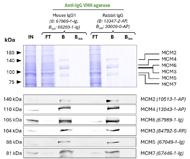
  • Co-IP of MCM complex via pulldown of MCM6 using 5 µg of anti-MCM6 antibodies and anti-rabbit IgG / anti-mouse IgG VHH agarose (mrIGa). All subunits of the 600 kDa heterohexameric complex are successfully precipitated using both a mouse IgG1 (67989-1-Ig) and a rabbit (13347-2-AP) MCM6 antibody, as shown by western blot anaylsis using subunit specific antibodies. Apparent molecular weights are provided. For Input (IN) and flow through (FT) fractions 1% was loaded. For bound (B) fraction and bound fraction of isotype control antibody (BISO), 20% was loaded. Product codes of PTG-antibodies are provided in parentheses. BISO is an isotype control used as negative control (ms1: 66260-1-Ig; rb: 30000-0-AP).

    Co-IP of MCM complex via pulldown of MCM6 using 5 µg of anti-MCM6 antibodies and anti-rabbit IgG / anti-mouse IgG VHH agarose (mrIGa). All subunits of the 600 kDa heterohexameric complex are successfully precipitated using both a mouse IgG1 (67989-1-Ig) and a rabbit (13347-2-AP) MCM6 antibody, as shown by western blot anaylsis using subunit specific antibodies. Apparent molecular weights are provided. For Input (IN) and flow through (FT) fractions 1% was loaded. For bound (B) fraction and bound fraction of isotype control antibody (BISO), 20% was loaded. Product codes of PTG-antibodies are provided in parentheses. BISO is an isotype control used as negative control (ms1: 66260-1-Ig; rb: 30000-0-AP).

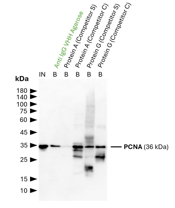
  • IP of PCNA using 5 µg of anti-PCNA antibody (IgG1, 60097-1-Ig) by anti-rabbit IgG/ anti-mouse IgG VHH agarose (mrIGa). For Western blot analysis the PCNA polyclonal antibody (10205-2-AP, 1:2000) was labeled using a conformation-specific HRP-conjugated secondary to avoid staining of heavy and light chain of the IP-antibody. In contrast to many competitor protein A and G beads, clean pulldown of PCNA by Proteintech anti-rabbit IgG/ anti-mouse IgG VHH agarose facilitates unambiguous identification of the target protein. Other beads show leaching of protein A or protein G fragments into the final IP fractions, which can lead to binding of the detection antibody and unspecific signals, as reported previously (Grant et al., 2019, Biiol Proced Online, doi: 10.1186/s12575-019-0095-z). For input (IN) and bound (B) fractions 1% and 30% were loaded, respectively.

    IP of PCNA using 5 µg of anti-PCNA antibody (IgG1, 60097-1-Ig) by anti-rabbit IgG/ anti-mouse IgG VHH agarose (mrIGa). For Western blot analysis the PCNA polyclonal antibody (10205-2-AP, 1:2000) was labeled using a conformation-specific HRP-conjugated secondary to avoid staining of heavy and light chain of the IP-antibody. In contrast to many competitor protein A and G beads, clean pulldown of PCNA by Proteintech anti-rabbit IgG/ anti-mouse IgG VHH agarose facilitates unambiguous identification of the target protein. Other beads show leaching of protein A or protein G fragments into the final IP fractions, which can lead to binding of the detection antibody and unspecific signals, as reported previously (Grant et al., 2019, Biiol Proced Online, doi: 10.1186/s12575-019-0095-z). For input (IN) and bound (B) fractions 1% and 30% were loaded, respectively.

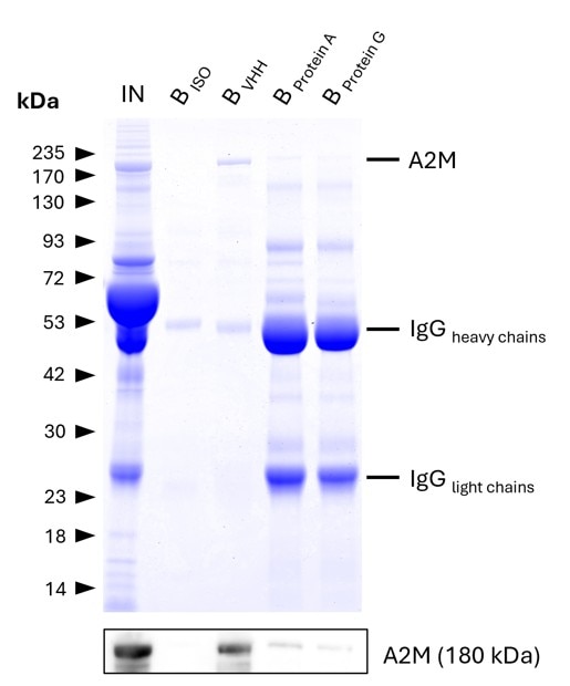
  • IP of Alpha-2-Macroglobulin (A2M) from human serum using anti-rabbit IgG / anti-mouse IgG VHH agarose (mrIGa) and comparison to protein A and G beads. IP was performed using 5 μg of rabbit anti-A2M IgG (13545-1-AP) in 1:10 diluted human serum. 0.5% and 6.25% of input (IN) and bound (B) fractions were analyzed by SDS-PAGE, respectively. 5 μg of rabbit isotype control antibody (BISO,  98136-1-RR) was used as negative control. The Coomassie-stained gel shows precipitation of A2M by anti-rabbit IgG / anti-mouse IgG VHH agarose and reveals its absence for protein A and G beads. This is likely due to human IgGs competing with the spiked IP-antibody for the latter beads. Results were verified using Western blot analysis, which detected A2M at approximately 180 kDa using an anti-A2M primary IgG (13545-1-AP, 1:5000).

    IP of Alpha-2-Macroglobulin (A2M) from human serum using anti-rabbit IgG / anti-mouse IgG VHH agarose (mrIGa) and comparison to protein A and G beads. IP was performed using 5 μg of rabbit anti-A2M IgG (13545-1-AP) in 1:10 diluted human serum. 0.5% and 6.25% of input (IN) and bound (B) fractions were analyzed by SDS-PAGE, respectively. 5 μg of rabbit isotype control antibody (BISO,  98136-1-RR) was used as negative control. The Coomassie-stained gel shows precipitation of A2M by anti-rabbit IgG / anti-mouse IgG VHH agarose and reveals its absence for protein A and G beads. This is likely due to human IgGs competing with the spiked IP-antibody for the latter beads. Results were verified using Western blot analysis, which detected A2M at approximately 180 kDa using an anti-A2M primary IgG (13545-1-AP, 1:5000).

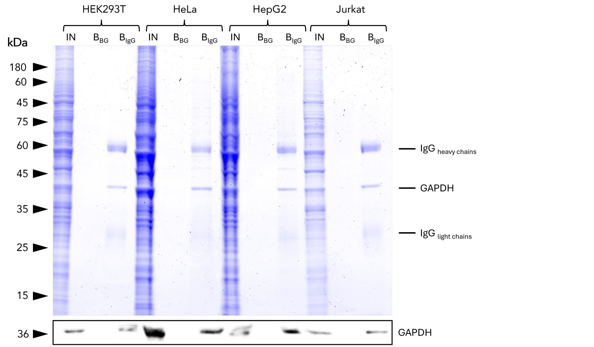
  • IP of GAPDH from common human cell lines using anti-rabbit IgG/ anti-mouse IgG VHH Agarose (mrIGa). IP was performed using 5 μg of rabbit anti-GAPDH IgG (10494-1-AP) with 1x10⁷ cells used per IP reaction. 1% and 25% of input (IN) and bound (B) fractions were analyzed by SDS-PAGE, respectively. Bound fractions without antibody spiked (BBG) are shown as background control. The Coomassie-stained gel shows precipitation of GAPDH by anti-rabbit IgG / anti-mouse IgG VHH agarose, what was verified using Western blot analysis using the anti-GAPDH mouse monoclonal antibody (60004-1-Ig).

    IP of GAPDH from common human cell lines using anti-rabbit IgG/ anti-mouse IgG VHH Agarose (mrIGa). IP was performed using 5 μg of rabbit anti-GAPDH IgG (10494-1-AP) with 1x10⁷ cells used per IP reaction. 1% and 25% of input (IN) and bound (B) fractions were analyzed by SDS-PAGE, respectively. Bound fractions without antibody spiked (BBG) are shown as background control. The Coomassie-stained gel shows precipitation of GAPDH by anti-rabbit IgG / anti-mouse IgG VHH agarose, what was verified using Western blot analysis using the anti-GAPDH mouse monoclonal antibody (60004-1-Ig).

ChromoTek anti-Rabbit IgG / anti-Mouse IgG VHH Agarose for Immunoprecipitation

anti-Rabbit / anti-Mouse IgG IP Beads is an affinity resin for IP of all subtypes of Rabbit and Mouse IgG. It consists of rabbit and mouse IgG specific VHHs (Nanobodies) coupled to Agarose beads.
Cat No. mrIGa

Reactivity

Rabbit IgG / Mouse IgG

Applications

IP, Co-IP

Host/Type

Alpaca, recombinant VHH

Conjugate

Agarose

Mouse IP, Rabbit IP, IgG IP, IgG Trap, Secondary VHH, VHH, Nanobody

Formats:  Agarose
Size/Concentration: 

-/ -


ご購入について

国内販売は「コスモ・バイオ株式会社」を通じて行っております。お見積り・ご注文はお近くの販売代理店へご連絡ください。


国内在庫・納期について

約2万点のプロテインテック製品をコスモバイオ社物流センター(国内)に在庫しています。国内在庫の有無はコスモバイオ社ホームページの「品番検索」でカタログ番号を検索して確認できます。


保証・サポートについて

テクニカルサポートまたはご購入後1年間の交換/補填対応を承ります。詳細はこちらをご覧ください。


Product Information

anti-Rabbit / anti-Mouse IgG IP Beads is an affinity resin for IP of all subtypes of Rabbit and Mouse IgG. It consists of rabbit and mouse IgG specific VHHs (Nanobodies) coupled to Agarose beads.

DescriptionReady to use VHH coupled Agarose Beads for Immunoprecipitation of all subtypes of Rabbit and Mouse IgG.

• Fast, reliable and efficient one-step Immunoprecipitation

• Ready-to-use

• Stable in harsh buffer conditions

• Suitable for downstream mass spec analysis

• Specific to all Rabbit and all Mouse IgG subtypes

• Low background

ApplicationsIP, Co-IP
Binding capacity/
ConjugateAgarose beads; bead size: ~ 90 µm (cross-linked 4 % agarose beads)
Elution bufferSDS Sample Buffer
Wash buffer compatibility/
TypeNanobody
ClassRecombinant - Animal free production
HostAlpaca
Affinity (KD)/
ImmunogenRabbit IgG / Mouse IgG
RRIDAB_3697100
Storage Buffer20% Ethanol
Storage Condition+4°C / do not freeze!
ShippingAmbient Temp
Size1 ml; 2 ml; 5 ml