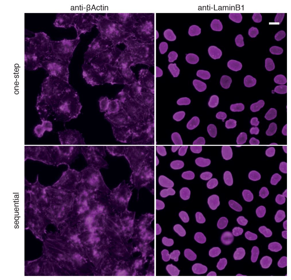
Validation Data Gallery
Product Information
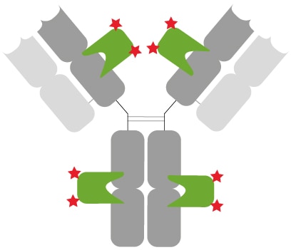
Nano-Secondary® anti-human IgG/anti-rabbit IgG, recombinant VHH is an anti-human IgG and anti-rabbit IgG specific secondary antibody. It consists of of a mixture of 2 Nanobodies that bind to human IgG and rabbit IgG with high affinity & specificity.
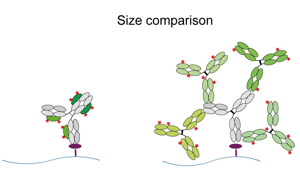
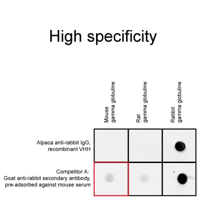
| Description | Nano-Secondary® alpaca anti-human IgG/anti-rabbit IgG, recombinant VHH reagent uses a novel class of anti-rabbit IgG and anti-human IgG specific antibodies. This secondary antibody product consists of Nanobodies that bind to rabbit IgG and human IgG with high affinity & specificity. Nano-Secondary reagents enable simultaneous incubation with primary antibodies, thus shortening your experimental. Due to their small size, Nano-Secondary reagents provide enhanced image resolution. No cross-reactivity against serum proteins from other species, details see below. |
| Applications | IF, WB, FC |
| Host | Alpaca |
| Species Reactivity | Rabbit, Human, Macaque No cross-reactivity to mouse, rat, sheep, goat, and guinea pig IgG |
| Target/Specificity | Fc-fragment of human IgG, Fab- and Fc-fragment of rabbit IgG (co-reactivity) |
| Physical State | Liquid |
| Conjugate | Alexa Fluor® 647 |
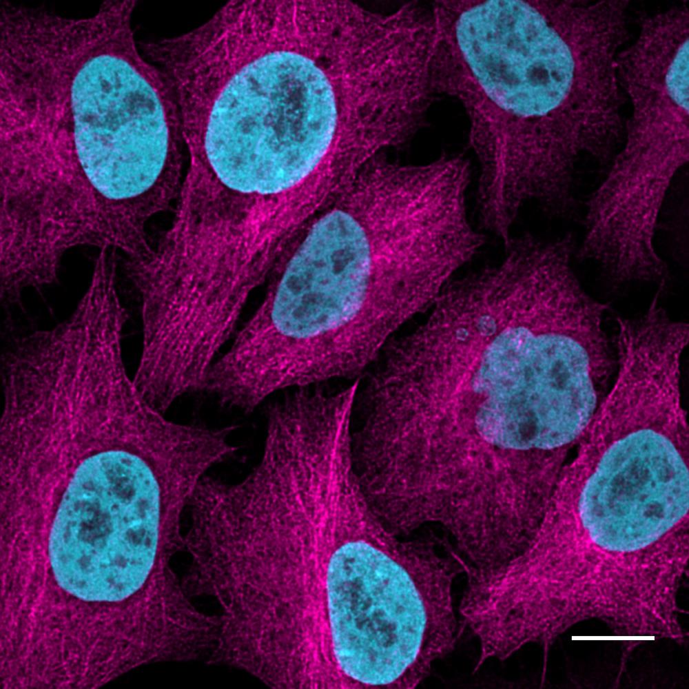
| Suggested Dilution | Immunofluorescence 1:1,000 Super-resolution microscopy 1:1,000 Western blot 1:1,000 |
| Purification Method | Recombinant expression, affinity purification IMAC |
| Type | Mixture of 2 monoclonal Nanobodies; Secondary Nanobody |
| Class | Recombinant |
| Format | Alpaca single domain antibody, monovalent |
| Cross-reactivity | No cross-reactivity to goat, guinea pig, mouse, rat, and sheep serum Cross-reactivity to macaque serum |
| Immunogen | Purified rabbit IgG |
| Clonality | Biclonal: mixture of 2 monoclonal Nanobodies |
| Clones | CTK0101 (VHH0244): Fc-fragment of human IgG, Fc-fragment of rabbit IgG CTK0102 (VHH0245): Fab-fragment of rabbit IgG |
| Affinity (KD) | CTK0101: KD = 0.2 nM, CTK0102: KD = 1.2 nM |
| RRID | AB_2827587 |
| Excitation / Emission | Excitation max: 650 nm, Emission max: 665 nm |
| Storage Buffer | 10 mM HEPES pH 7.0, 500 mM NaCl, 5 mM EDTA, Preservative: 0.09 % Sodium azide |
| Storage Condition | Store at -20°C short term or -80°C long term. Aliquot upon delivery. Avoid freeze-thaw cycles. |
| Size | 10 µL; 100 µL |
| Note | This product is for research use only, not for diagnostic or therapeutic use |
Documentation
| SDS |
|---|
| srbAF647-1_SDS_Nano-Secondary® alpaca anti-human IgG, anti-rabbit IgG, recombinant VHH, Alexa Fluor® 647 [CTK0101, CTK0102] (EN) |
| Datasheet |
|---|
| Nano-Secondary® anti-human IgG/anti-rabbit IgG, recombinant VHH, Alexa Fluor® 647 [CTK0101, CTK0102] Datasheet |
| Application note |
|---|
| One-step incubation with Nano-Secondaries (PDF) |
| Blocking recommendation for immunostaining with Nano-Secondaries (PDF) |
| Brochure |
|---|
| Chromotek nanobodies brochure (PDF) |
Publications
| Application | Title |
|---|---|
Biosens Bioelectron 3D co-culture of macrophages and fibroblasts in a sessile drop array for unveiling the role of macrophages in skin wound-healing | |
Vet Microbiol NDV inhibited IFN-β secretion through impeding CHCHD10-mediated mitochondrial fusion to promote viral proliferation | |
Nat Methods Spatial single-cell mass spectrometry defines zonation of the hepatocyte proteome |

