Validation Data Gallery

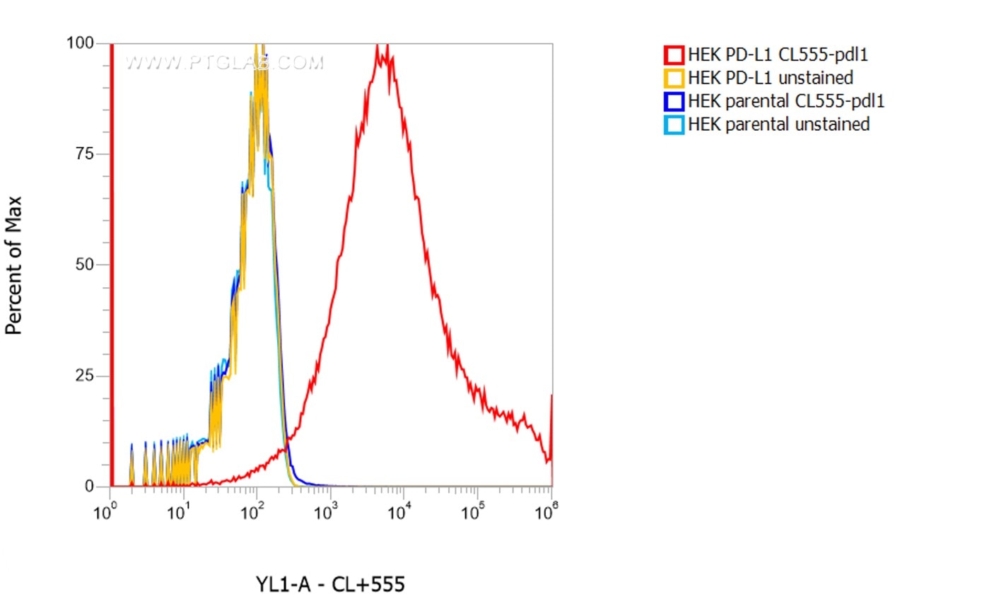
  • 1X10^6 HEK PD-L1 transient transfected cells (red) and HEK parental cells were surface stained with 0.5 µg CoraLite® Plus 555 conjugated-PD-L1 VHH (CL555-pdlo). Cells were not fixed.

    1X10^6 HEK PD-L1 transient transfected cells (red) and HEK parental cells were surface stained with 0.5 µg CoraLite® Plus 555 conjugated-PD-L1 VHH (CL555-pdlo). Cells were not fixed.

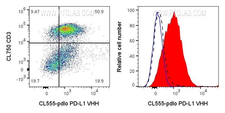
  • Left: 1x10^6 PHA-treated (3d) human PBMCs were surface stained with 0.125ug CoraLite® Plus 555 conjugated PD-L1 VHH (CL555-pdlo) and CL750 CD3 (CL750-65151). Right: 1x10^6 PHA-treated (3d) human PBMCs were surface stained with 0.125ug CoraLite® Plus 555 conjugated PD-L1 VHH (CL555-pdlo) (red) or unstained (blue). 1X10^6 untreated human PBMCs were surface stained with 0.125ug CoraLite® Plus 555 conjugated PD-L1 VHH (CL555-pdlo) (black dashed). Cells were treated with FC Receptor Block prior to staining. Cells were not fixed. Lymphocytes were gated.

    Left: 1x10^6 PHA-treated (3d) human PBMCs were surface stained with 0.125ug CoraLite® Plus 555 conjugated PD-L1 VHH (CL555-pdlo) and CL750 CD3 (CL750-65151). Right: 1x10^6 PHA-treated (3d) human PBMCs were surface stained with 0.125ug CoraLite® Plus 555 conjugated PD-L1 VHH (CL555-pdlo) (red) or unstained (blue). 1X10^6 untreated human PBMCs were surface stained with 0.125ug CoraLite® Plus 555 conjugated PD-L1 VHH (CL555-pdlo) (black dashed). Cells were treated with FC Receptor Block prior to staining. Cells were not fixed. Lymphocytes were gated.

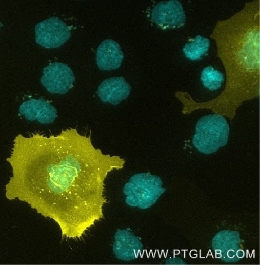
  • A431 cells transfected with human PD-L1 were immunostained with CoraLite® Plus 555 conjugated-PD-L1 VHH (CL555-pdlo) (1:1000, yellow). Nuclei were stained with DAPI (cyan). Epifluorescence images were acquired with a 20x objective and post-processed.
 
Comment: Immunostaining with CL555-pdlo can be performed live or after formaldehyde fixation.

    A431 cells transfected with human PD-L1 were immunostained with CoraLite® Plus 555 conjugated-PD-L1 VHH (CL555-pdlo) (1:1000, yellow). Nuclei were stained with DAPI (cyan). Epifluorescence images were acquired with a 20x objective and post-processed. Comment: Immunostaining with CL555-pdlo can be performed live or after formaldehyde fixation.

ChromoTek CoraLite® Plus 555-conjugated PD-L1 VHH

CL555-pdlo targets PD-L1 in FC and IF applications and shows reactivity with Human samples.
Cat No. CL555-pdlo

Host/Type

Alpaca VHH

Reactivity

Human

Applications

FC, IF

Conjugate

CoraLite® Plus 555

PD-L1-Nanobody, PD-L1-Single domain antibody (sdAb), B7 homolog 1, B7-H1, CD274, programmed cell death 1 ligand 1, PDL1, PD-L1

Size/Concentration: 

-/ -


ご購入について

国内販売は「コスモ・バイオ株式会社」を通じて行っております。お見積り・ご注文はお近くの販売代理店へご連絡ください。


国内在庫・納期について

約2万点のプロテインテック製品をコスモバイオ社物流センター(国内)に在庫しています。国内在庫の有無はコスモバイオ社ホームページの「品番検索」でカタログ番号を検索して確認できます。


保証・サポートについて

テクニカルサポートまたはご購入後1年間の交換/補填対応を承ります。詳細はこちらをご覧ください。


Product Information

CL555-pdlo targets PD-L1 in FC and IF applications and shows reactivity with Human samples.

ApplicationsFC, IF
ReactivityHuman
ConjugateCoraLite® Plus 555
TypeNanobody
ClassRecombinant
HostAlpaca
AffinityPicomolar range, below the assay limit (biolayer interferometry)
Molecular Weight13.887 kDa
FormLiquid
Excitation/Emission Maxima Wavelengths554 nm / 570 nm
RRIDAB_3661835
Storage Buffer500 mM NaCl, 10 mM HEPES pH 7.0, 5 mM EDTA, 0.09% sodium azide
Storage ConditionStore at -20°C
BackgroundPD-L1, also known as CD274 or B7H1, stands for programmed cell death ligand 1. It is a type I transmembrane protein that is thought to repress immune responses by binding to its receptor (PD1), thus inhibiting T-cell activation, proliferation, and cytokine production. It contains V-like and C-like immunoglobulin domains. PD-L1 expression is regulated by various cytokines, such as TNF-α or LPS (ISSN: 1848-7718). Increased expression of this protein in certain types of cancers, e.g., renal cell carcinoma or colon cancer, correlates with poor prognosis. D-L1 is critical for the induction and maintenance of immune self-tolerance during infection or inflammation in normal tissues. The interaction of PD-L1 and its receptors is responsible for preventing auto-immune phenotypes and balancing the overall immune response in situations such as pregnancy or tissue allografts. The interaction between PD-L1 and PD-1 or B7.1 starts an inhibitory signaling cascade, which results in the decreased proliferation of antigen-specific T-cells and increased survival of regulatory T-cells (PMID: 15240681).