WB Figures
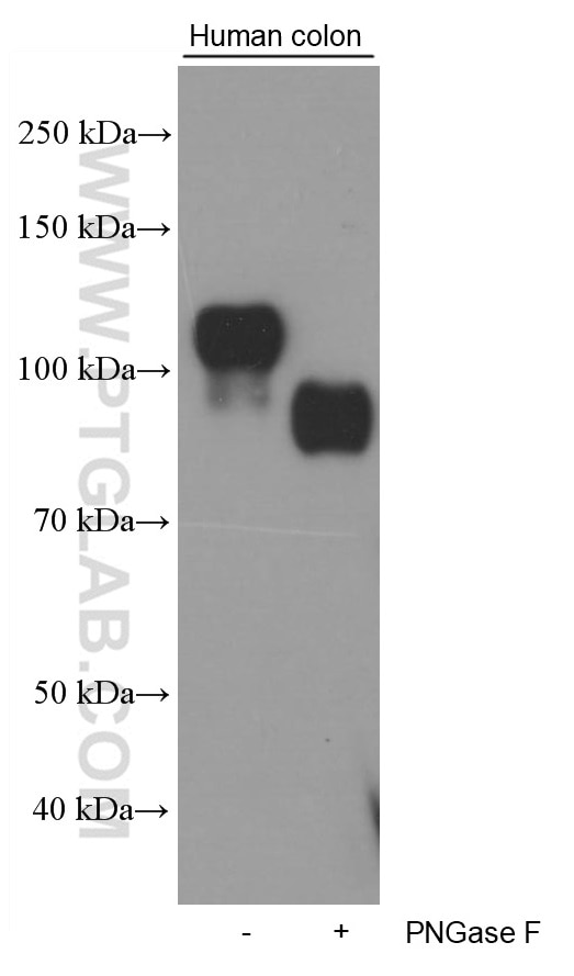
WB analysis of human colon using 60351-1-Ig (same clone as 60351-1-PBS)
Untreated and PNGase F-treated lysates of human colon tissue were subjected to SDS PAGE followed by western blot with 60351-1-Ig (Cadherin-17 antibody) at dilution of 1:5000 incubated at room temperature for 1.5 hours. PNGase F was obtained from Atagenix (cat.NO. ata808). This data was developed using the same antibody clone with 60351-1-PBS in a different storage buffer formulation.
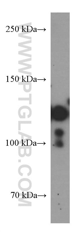
WB analysis of pig colon using 60351-1-Ig (same clone as 60351-1-PBS)
pig colon tissue were subjected to SDS PAGE followed by western blot with 60351-1-Ig (Cadherin-17 antibody) at dilution of 1:2000 incubated at room temperature for 1.5 hours. This data was developed using the same antibody clone with 60351-1-PBS in a different storage buffer formulation.
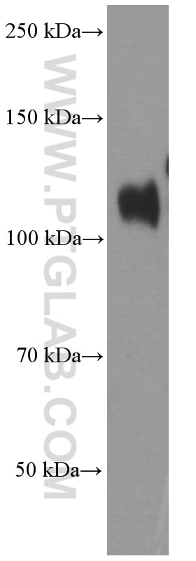
WB analysis of human colon using 60351-1-Ig (same clone as 60351-1-PBS)
human colon tissue were subjected to SDS PAGE followed by western blot with 60351-1-Ig (Cadherin-17 antibody) at dilution of 1:20000 incubated at room temperature for 1.5 hours. This data was developed using the same antibody clone with 60351-1-PBS in a different storage buffer formulation.
IHC Figures
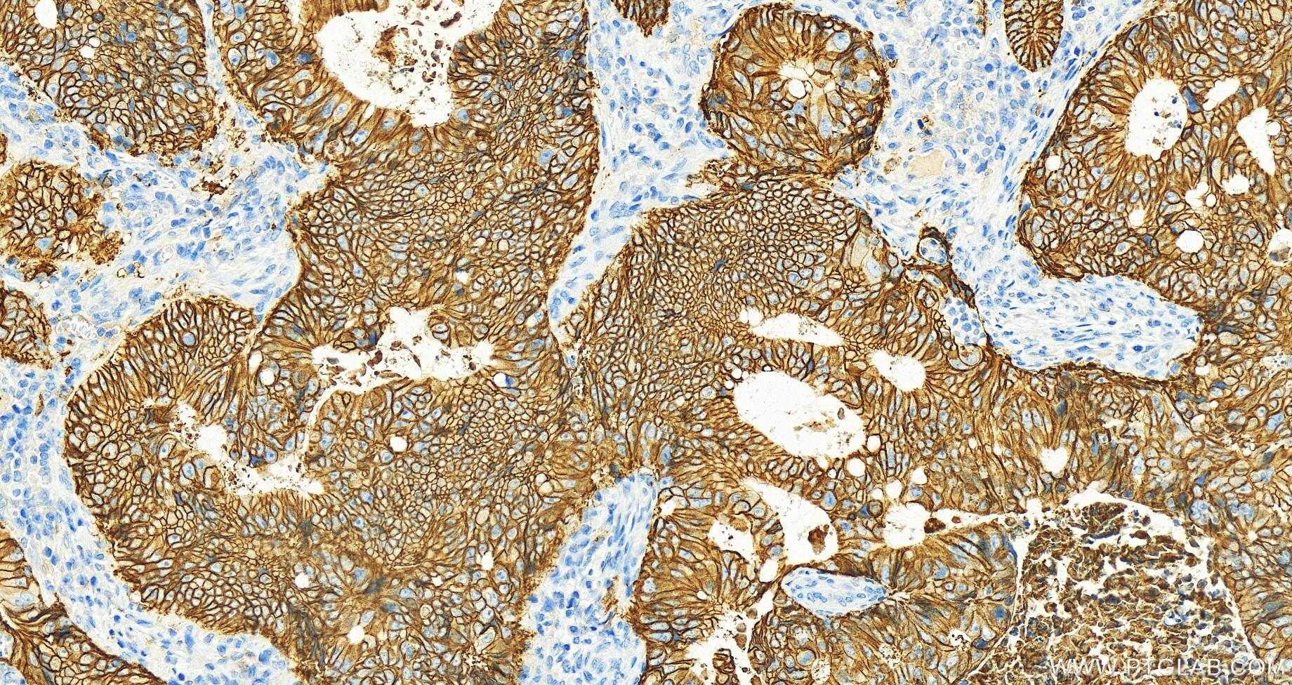
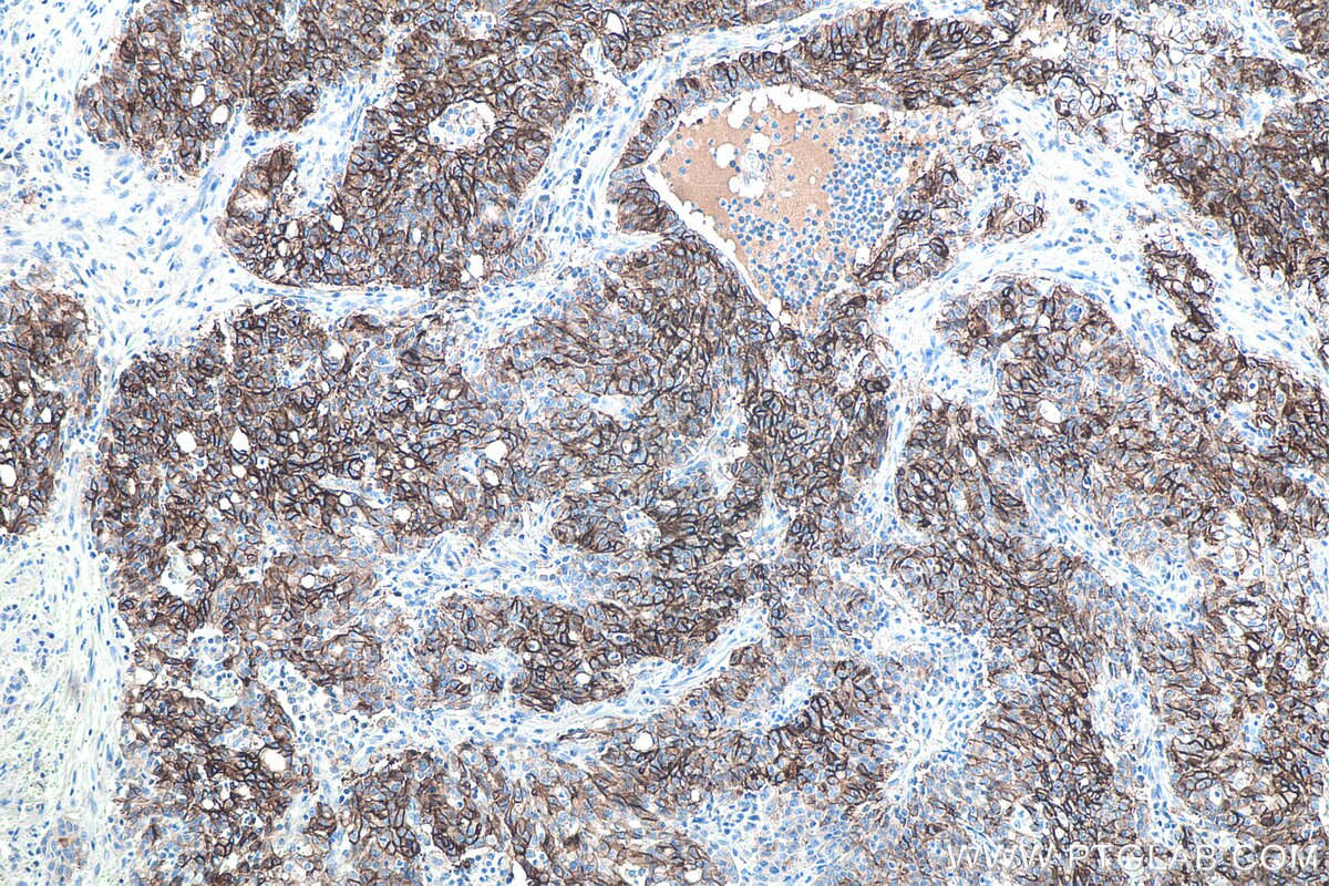
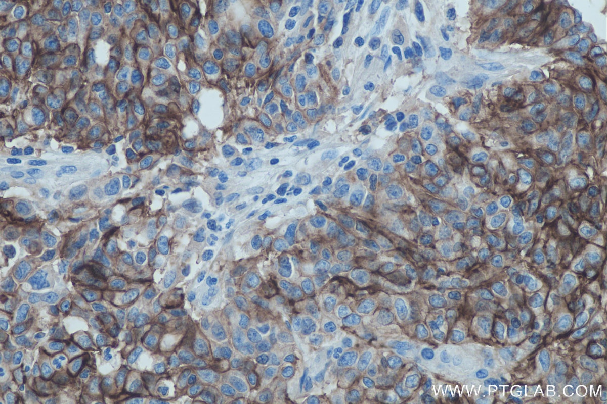
IHC staining of human colon cancer using 60351-1-Ig (same clone as 60351-1-PBS)
Immunohistochemical analysis of paraffin-embedded colon cancer slide using 60351-1-Ig (Cadherin-17 antibody) at dilution of 1:1000 (under 20x lens). Heat mediated antigen retrieval with Tris-EDTA buffer (pH 9.0). This data was developed using the same antibody clone with 60351-1-PBS in a different storage buffer formulation.
IHC staining of human colon cancer using 60351-1-Ig (same clone as 60351-1-PBS)
Immunohistochemical analysis of paraffin-embedded colon cancer slide using 60351-1-Ig (Cadherin-17 antibody) at dilution of 1:1000 (under 20x lens). Heat mediated antigen retrieval with Tris-EDTA buffer (pH 9.0). This data was developed using the same antibody clone with 60351-1-PBS in a different storage buffer formulation.
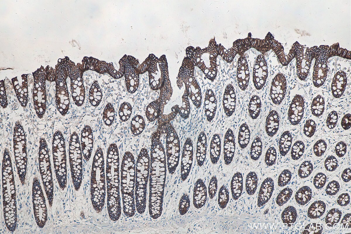
IHC staining of human colon using 60351-1-Ig (same clone as 60351-1-PBS)
Immunohistochemical analysis of paraffin-embedded human colon tissue slide using 60351-1-Ig (Cadherin-17 antibody) at dilution of 1:32000 (under 10x lens). Heat mediated antigen retrieval with Tris-EDTA buffer (pH 9.0). This data was developed using the same antibody clone with 60351-1-PBS in a different storage buffer formulation.
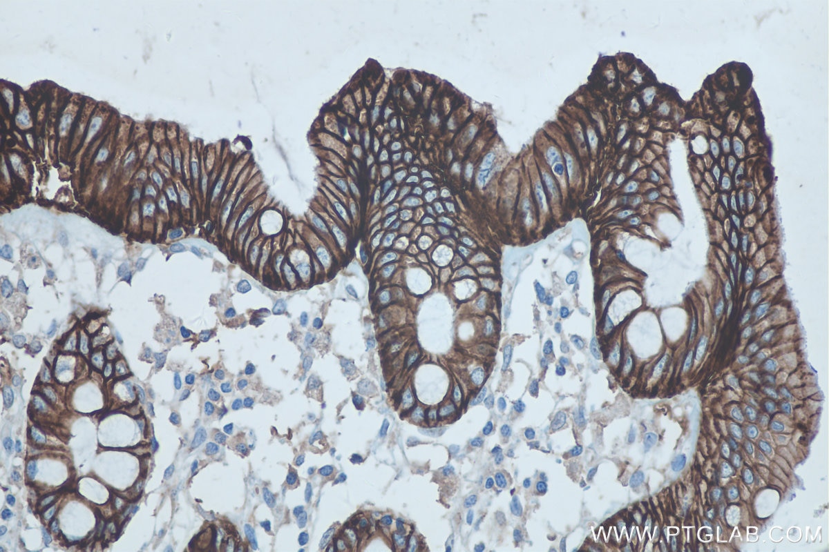
IHC staining of human colon using 60351-1-Ig (same clone as 60351-1-PBS)
Immunohistochemical analysis of paraffin-embedded human colon tissue slide using 60351-1-Ig (Cadherin-17 antibody) at dilution of 1:32000 (under 40x lens). Heat mediated antigen retrieval with Tris-EDTA buffer (pH 9.0). This data was developed using the same antibody clone with 60351-1-PBS in a different storage buffer formulation.
IHC staining of human stomach cancer using 60351-1-Ig (same clone as 60351-1-PBS)
Immunohistochemical analysis of paraffin-embedded human stomach cancer tissue slide using 60351-1-Ig (Cadherin-17 antibody) at dilution of 1:16000 (under 10x lens). Heat mediated antigen retrieval with Tris-EDTA buffer (pH 9.0). This data was developed using the same antibody clone with 60351-1-PBS in a different storage buffer formulation.
IHC staining of human stomach cancer using 60351-1-Ig (same clone as 60351-1-PBS)
Immunohistochemical analysis of paraffin-embedded human stomach cancer tissue slide using 60351-1-Ig (Cadherin-17 antibody) at dilution of 1:16000 (under 40x lens). Heat mediated antigen retrieval with Tris-EDTA buffer (pH 9.0). This data was developed using the same antibody clone with 60351-1-PBS in a different storage buffer formulation.
IHC staining of human stomach cancer using 60351-1-Ig (same clone as 60351-1-PBS)
Immunohistochemical analysis of paraffin-embedded human stomach cancer tissue slide using 60351-1-Ig (Cadherin-17 antibody) at dilution of 1:8000. Heat mediated antigen retrieval with Tris-EDTA buffer (pH 9.0). This data was developed using the same antibody clone with 60351-1-PBS in a different storage buffer formulation.
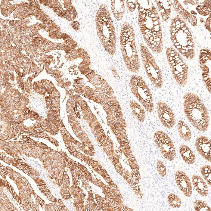
IHC staining of human colorectal cancer using 60351-1-Ig (same clone as 60351-1-PBS)
Immunohistochemical analysis of paraffin-embedded human colorectal cancer tissue slide using 60351-1-Ig (Cadherin-17 antibody) at dilution of 1:8000. Heat mediated antigen retrieval with Tris-EDTA buffer (pH 9.0). This data was developed using the same antibody clone with 60351-1-PBS in a different storage buffer formulation.
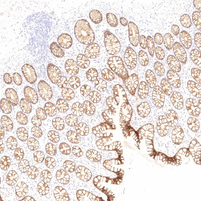
IHC staining of human large intestine using 60351-1-Ig (same clone as 60351-1-PBS)
Immunohistochemical analysis of paraffin-embedded human large intestine tissue slide using 60351-1-Ig (Cadherin-17 antibody) at dilution of 1:8000. Heat mediated antigen retrieval with Tris-EDTA buffer (pH 9.0). This data was developed using the same antibody clone with 60351-1-PBS in a different storage buffer formulation.
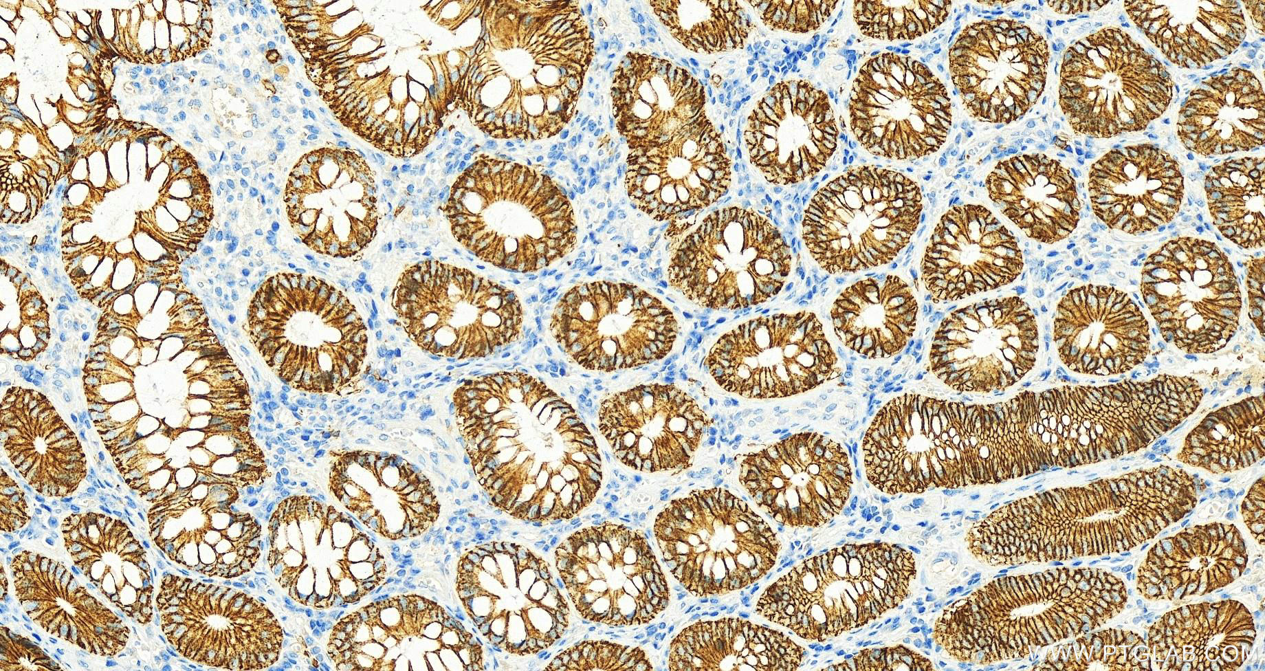
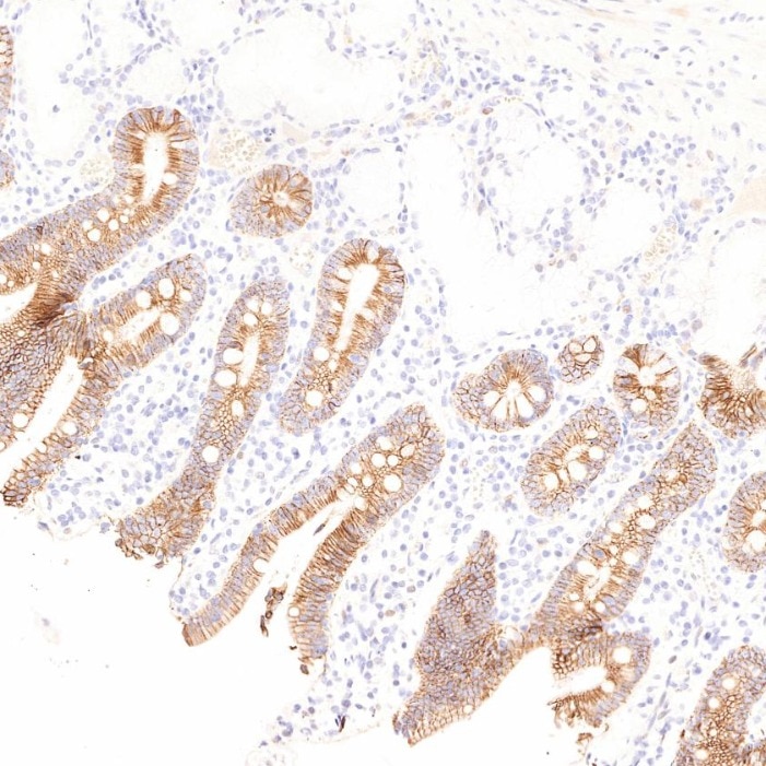
IHC staining of human small intestine using 60351-1-Ig (same clone as 60351-1-PBS)
Immunohistochemical analysis of paraffin-embedded human small intestine tissue slide using 60351-1-Ig (Cadherin-17 antibody) at dilution of 1:8000. Heat mediated antigen retrieval with Tris-EDTA buffer (pH 9.0). This data was developed using the same antibody clone with 60351-1-PBS in a different storage buffer formulation.
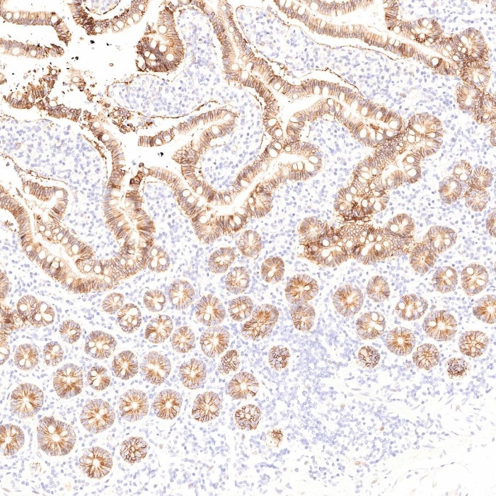
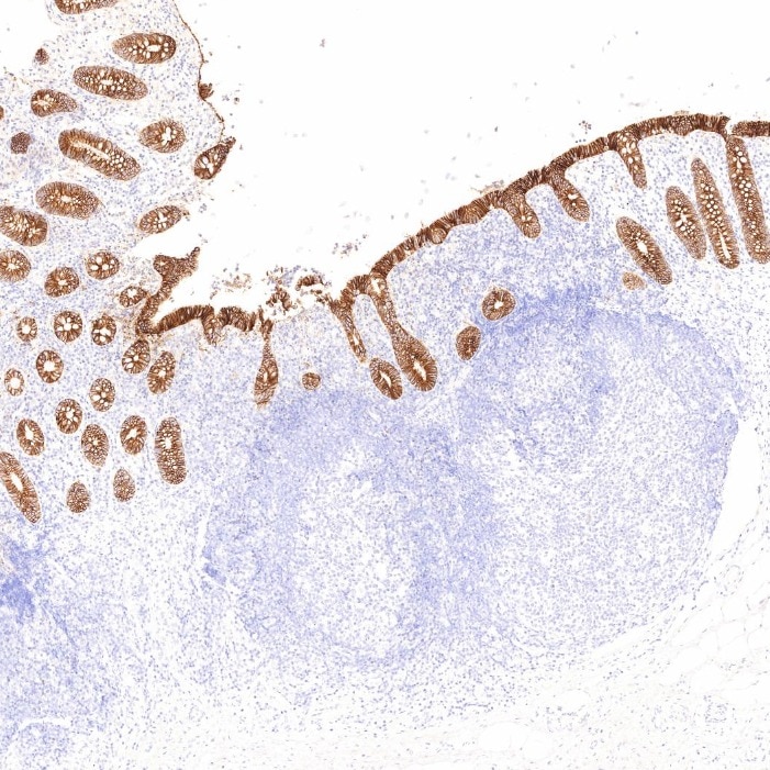
IHC staining of human appendicitis using 60351-1-Ig (same clone as 60351-1-PBS)
Immunohistochemical analysis of paraffin-embedded human appendicitis tissue slide using 60351-1-Ig (Cadherin-17 antibody) at dilution of 1:8000. Heat mediated antigen retrieval with Tris-EDTA buffer (pH 9.0). This data was developed using the same antibody clone with 60351-1-PBS in a different storage buffer formulation.
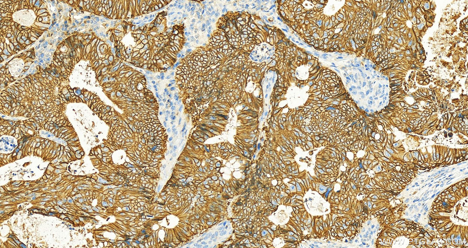
IHC staining of human colon cancer using 60351-1-Ig (same clone as 60351-1-PBS)
Immunohistochemical analysis of paraffin-embedded colon cancer slide using 60351-1-Ig (Cadherin-17 antibody) at dilution of 1:1000 (under 20x lens). Heat mediated antigen retrieval with Tris-EDTA buffer (pH 9.0). This data was developed using the same antibody clone with 60351-1-PBS in a different storage buffer formulation.
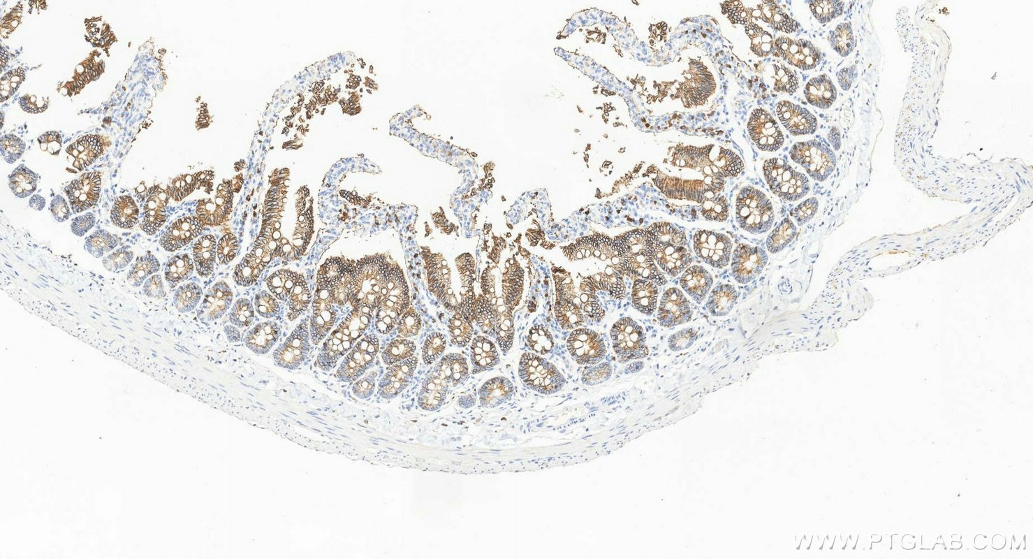
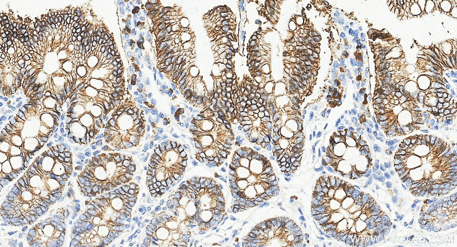
IHC staining of rat small intestine using 60351-1-Ig (same clone as 60351-1-PBS)
Immunohistochemical analysis of paraffin-embedded rat small intestine tissue slide using 60351-1-Ig (Cadherin-17 antibody) at dilution of 1:5000 (under 10x lens). Heat mediated antigen retrieval with Tris-EDTA buffer (pH 9.0). This data was developed using the same antibody clone with 60351-1-PBS in a different storage buffer formulation.
IHC staining of rat small intestine using 60351-1-Ig (same clone as 60351-1-PBS)
Immunohistochemical analysis of paraffin-embedded rat small intestine tissue slide using 60351-1-Ig (Cadherin-17 antibody) at dilution of 1:5000 (under 40x lens). Heat mediated antigen retrieval with Tris-EDTA buffer (pH 9.0). This data was developed using the same antibody clone with 60351-1-PBS in a different storage buffer formulation.
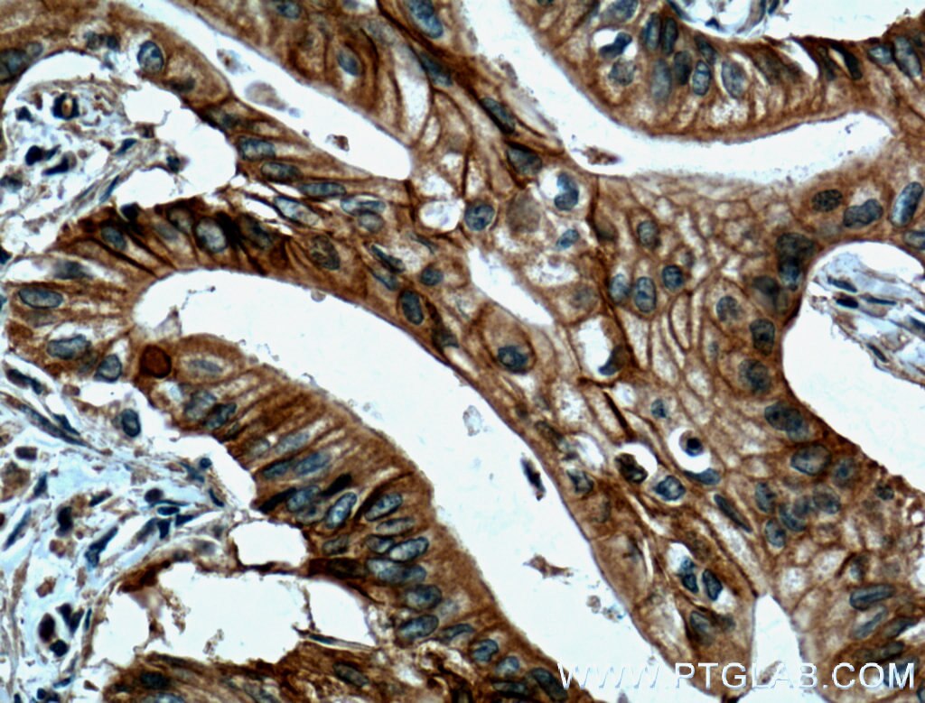
IHC staining of human pancreas cancer using 60351-1-Ig (same clone as 60351-1-PBS)
Immunohistochemical analysis of paraffin-embedded human pancreas cancer tissue slide using 60351-1-Ig (Cadherin-17 antibody) at dilution of 1:800 (under 40x lens. Heat mediated antigen retrieval with Tris-EDTA buffer (pH 9.0). This data was developed using the same antibody clone with 60351-1-PBS in a different storage buffer formulation.
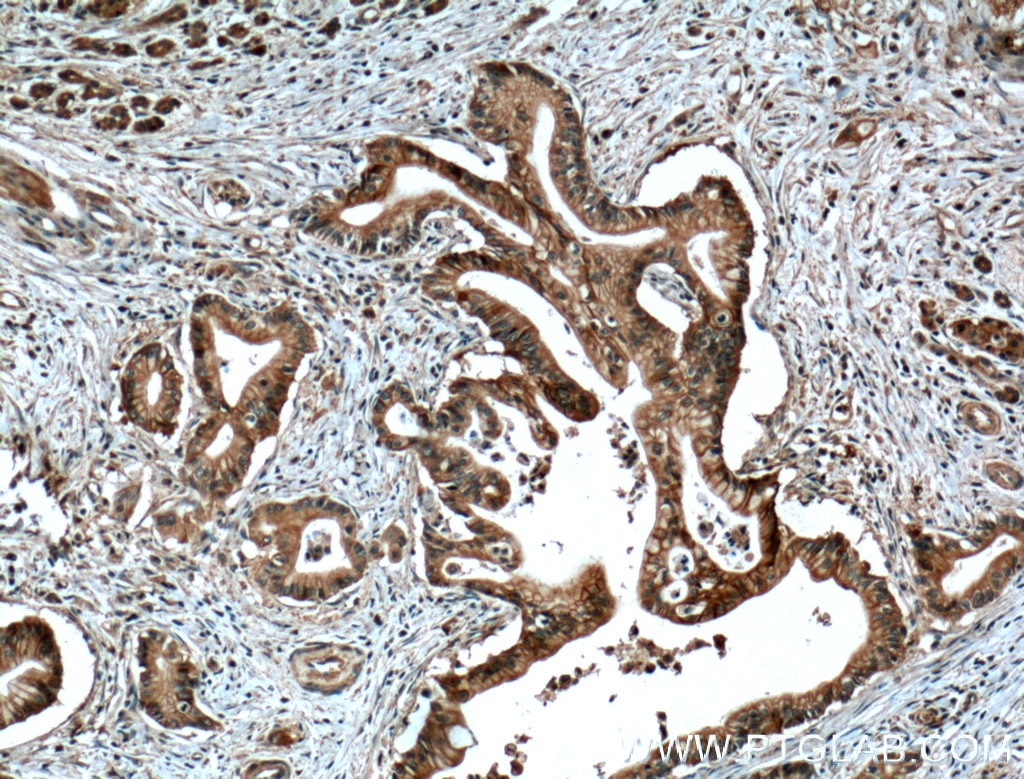
IHC staining of human pancreas cancer using 60351-1-Ig (same clone as 60351-1-PBS)
Immunohistochemical analysis of paraffin-embedded human pancreas cancer tissue slide using 60351-1-Ig (Cadherin-17 antibody) at dilution of 1:800 (under 10x lens. Heat mediated antigen retrieval with Tris-EDTA buffer (pH 9.0). This data was developed using the same antibody clone with 60351-1-PBS in a different storage buffer formulation.
IF-P Figures
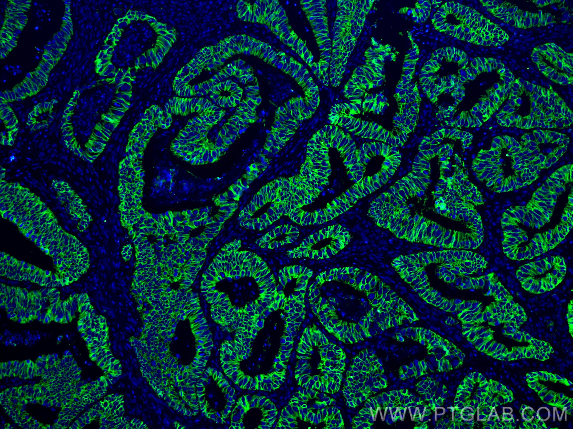
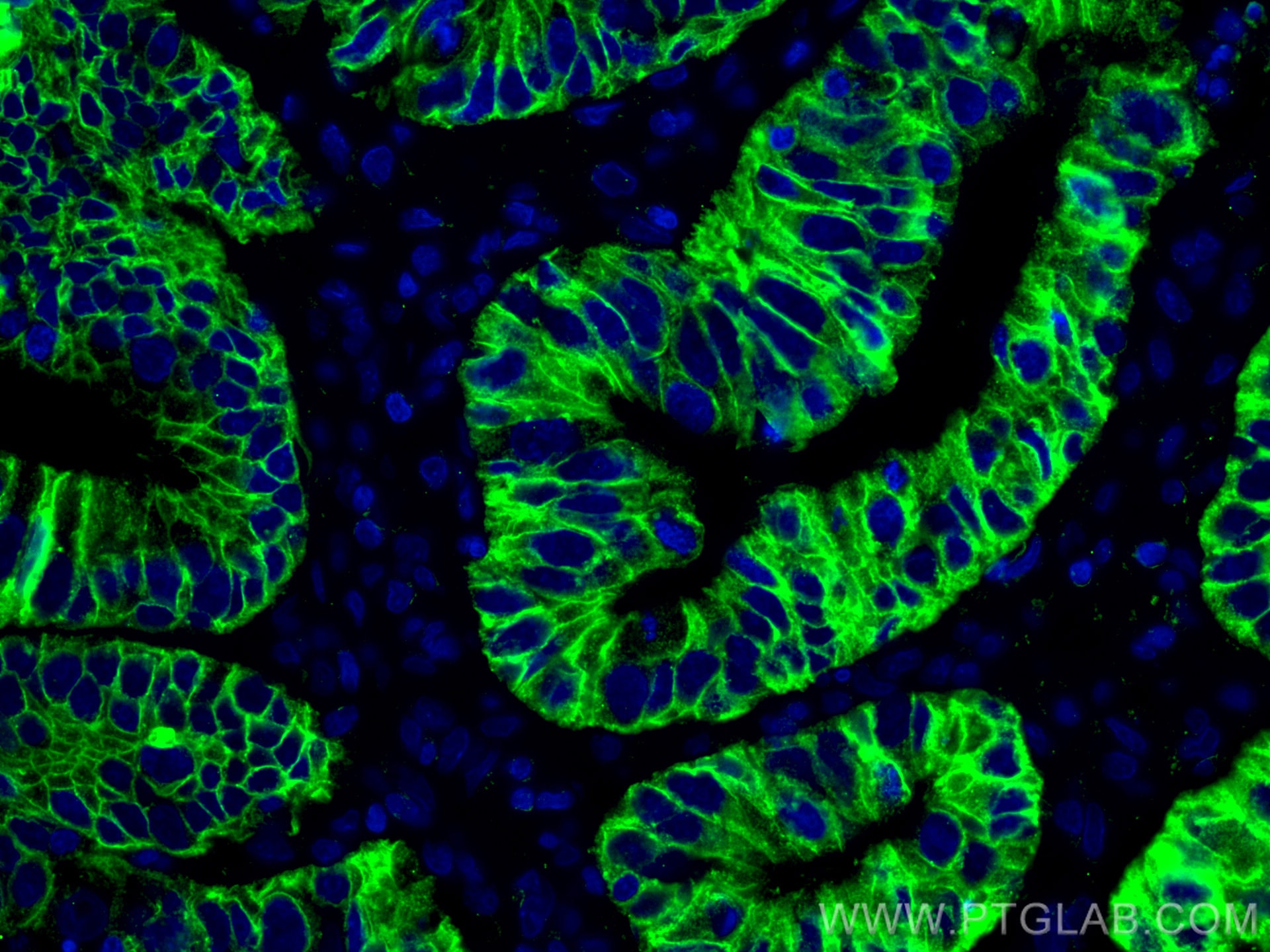
IF Staining of human colon cancer using 60351-1-Ig (same clone as 60351-1-PBS)
Immunofluorescent analysis of (4% PFA) fixed human colon cancer tissue using Cadherin-17 antibody (60351-1-Ig, Clone: 3B10A9 ) at dilution of 1:400 and CoraLite®488-Conjugated AffiniPure Goat Anti-Mouse IgG(H+L). This data was developed using the same antibody clone with 60351-1-PBS in a different storage buffer formulation.
IF Staining of human colon cancer using 60351-1-Ig (same clone as 60351-1-PBS)
Immunofluorescent analysis of (4% PFA) fixed human colon cancer tissue using Cadherin-17 antibody (60351-1-Ig, Clone: 3B10A9 ) at dilution of 1:400 and CoraLite®488-Conjugated AffiniPure Goat Anti-Mouse IgG(H+L). This data was developed using the same antibody clone with 60351-1-PBS in a different storage buffer formulation.