Validation Data Gallery
Tested Applications
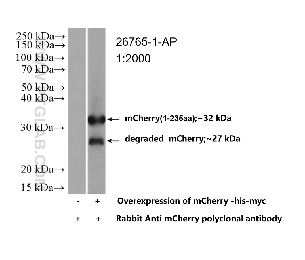
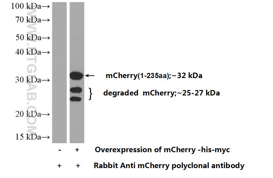
| Positive WB detected in | Transfected HEK-293 cells, Recombinant protein |
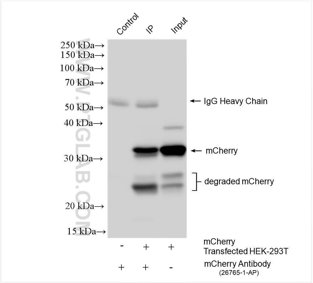
| Positive IP detected in | Transfected HEK-293T cells |
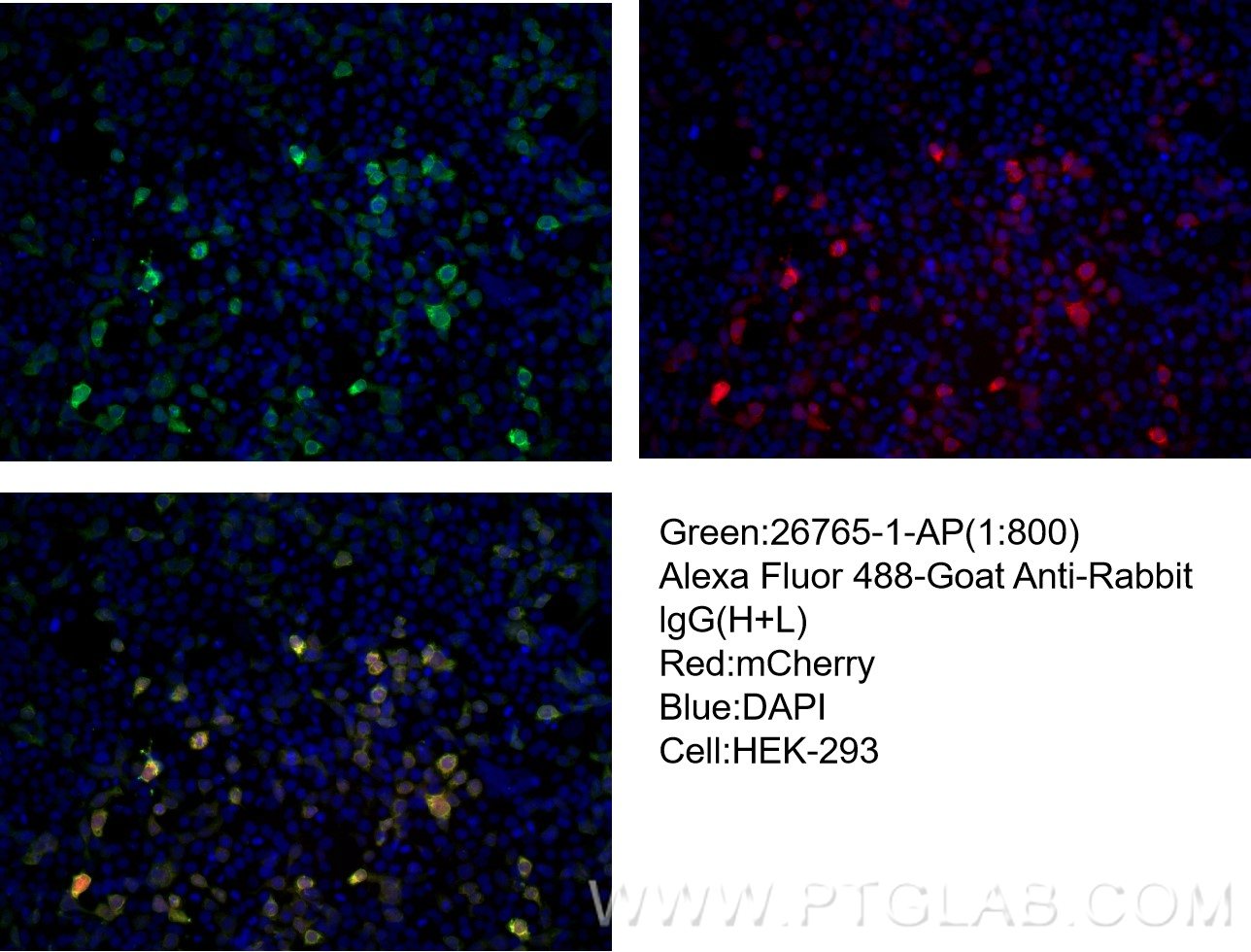
| Positive IF/ICC detected in | Transfected HEK-293 cells |
Recommended dilution
| Application | Dilution |
|---|---|
| Western Blot (WB) | WB : 1:1000-1:4000 |
| Immunoprecipitation (IP) | IP : 0.5-4.0 ug for 1.0-3.0 mg of total protein lysate |
| Immunofluorescence (IF)/ICC | IF/ICC : 1:400-1:1600 |
| It is recommended that this reagent should be titrated in each testing system to obtain optimal results. | |
| Sample-dependent, Check data in validation data gallery. | |
Published Applications
| WB | See 68 publications below |
| IHC | See 4 publications below |
| IF | See 41 publications below |
| IP | See 11 publications below |
| CoIP | See 4 publications below |
Product Information
26765-1-AP targets mCherry in WB, IHC, IF/ICC, IP, CoIP, ELISA applications and shows reactivity with recombinant protein samples.
| Tested Reactivity | recombinant protein |
| Cited Reactivity | human, mouse, rabbit, monkey, zebrafish, yeast, plant, d. punctatus |
| Host / Isotype | Rabbit / IgG |
| Class | Polyclonal |
| Type | Antibody |
| Immunogen |
CatNo: Ag25320 Product name: Recombinant mCherry protein Source: e coli.-derived, PGEX-4T Tag: GST Sequence: VSKGEEDNMAIIKEFMRFKVHMEGSVNGHEFEIEGEGEGRPYEGTQTAKLKVTKGGPLPFAWDILSPQFMYGSKAYVKHPADIPDYLKLSFPEGFKWERVMNFEDGGVVTVTQDSSLQDGEFIYKVKLRGTNFPSDGPVMQKKTMGWEASSERMYPEDGALKGEIKQRLKLKDGGHYDAEVKTTYKAKKPVQLPGAYNVNIKLDITSHNEDYTIVEQYERAEGRHSTGGMDELYK 相同性解析による交差性が予測される生物種 |
| Full Name | mCherry |
| Calculated molecular weight | 27 kDa |
| Gene Symbol | |
| Gene ID (NCBI) | |
| RRID | AB_2876881 |
| Conjugate | Unconjugated |
| Form | |
| Form | Liquid |
| Purification Method | Antigen affinity purification |
| Storage Buffer | PBS with 0.02% sodium azide and 50% glycerol{{ptg:BufferTemp}}7.3 |
| Storage Conditions | Store at -20°C. Stable for one year after shipment. Aliquoting is unnecessary for -20oC storage. |
Background Information
Red fluorescent proteins (RFPs) is a collective term referring to a heterogenous group of red chromophore-carrying proteins, originating from various species and forming different protein lineages.
The original RFP (dsRed) is a 225 amino acid fluorescent protein (25.9 kDa) derived from Discosoma sp.. It emits red light with a peak wavelength of 593 nm upon excitation by green light (excitation peak at 558 nm).
When fused with other proteins, RFP serves as a versatile reporter protein e.g. for quantifying expression levels or facilitates visualization of subcellular localization through fluorescence microscopy.
This antibody is a rabbit polyclonal antibody raised against mCherry. It can be used to detect mCherry, dsRed, tdTomato, and mScarlet.
Protocols
| Product Specific Protocols | |
|---|---|
| IF protocol for mCherry antibody 26765-1-AP | Download protocol |
| IP protocol for mCherry antibody 26765-1-AP | Download protocol |
| WB protocol for mCherry antibody 26765-1-AP | Download protocol |
| Standard Protocols | |
|---|---|
| Click here to view our Standard Protocols |
Publications
| Species | Application | Title |
|---|---|---|
Nat Struct Mol Biol Aurora kinase A-mediated phosphorylation triggers structural alteration of Rab1A to enhance ER complexity during mitosis | ||
Nat Commun Stalled translation by mitochondrial stress upregulates a CNOT4-ZNF598 ribosomal quality control pathway important for tissue homeostasis | ||
J Extracell Vesicles Identification of the SNARE complex that mediates the fusion of multivesicular bodies with the plasma membrane in exosome secretion | ||
Cell Death Differ RING1 dictates GSDMD-mediated inflammatory response and host susceptibility to pathogen infection |


