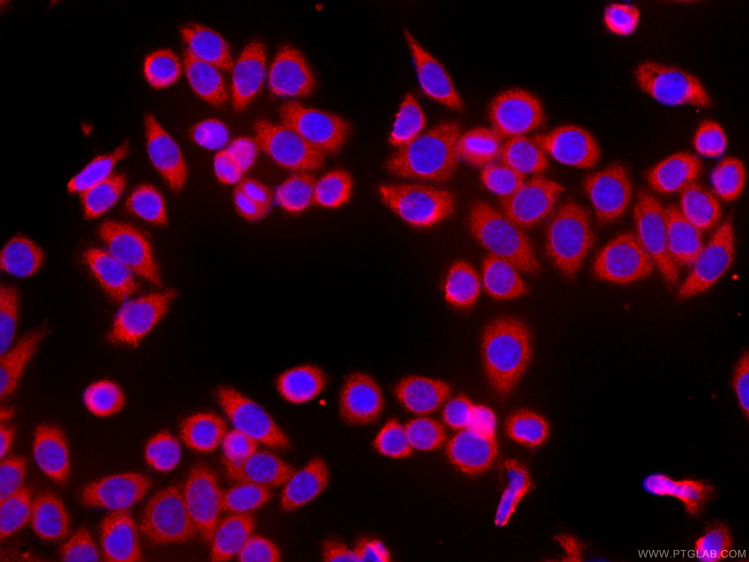
CoraLite®594-conjugated HSPH1 Monoclonal antibody
HSPH1 Monoclonal Antibody for IF
Host / Isotype
Mouse / IgG1
Reactivity
Human
Applications
IF
Conjugate
CoraLite®594 Fluorescent Dye
CloneNo.
3H5A5
Cat no : CL594-66723
Synonyms
Validation Data Gallery
Tested Applications
| Positive IF detected in | HeLa cells |
Recommended dilution
| Application | Dilution |
|---|---|
| Immunofluorescence (IF) | IF : 1:50-1:500 |
| Sample-dependent, check data in validation data gallery | |
Product Information
CL594-66723 targets HSPH1 in IF applications and shows reactivity with Human samples.
| Tested Reactivity | Human |
| Host / Isotype | Mouse / IgG1 |
| Class | Monoclonal |
| Type | Antibody |
| Immunogen | HSPH1 fusion protein Ag27521 相同性解析による交差性が予測される生物種 |
| Full Name | heat shock 105kDa/110kDa protein 1 |
| Calculated molecular weight | 858 aa, 97 kDa |
| Observed molecular weight | 110 kDa |
| GenBank accession number | BC037553 |
| Gene symbol | HSPH1 |
| Gene ID (NCBI) | 10808 |
| RRID | AB_2883618 |
| Conjugate | CoraLite®594 Fluorescent Dye |
| Excitation/Emission maxima wavelengths | 588 nm / 604 nm |
| Form | Liquid |
| Purification Method | Protein G purification |
| Storage Buffer | PBS with 50% Glycerol, 0.05% Proclin300, 0.5% BSA, pH 7.3. |
| Storage Conditions | Store at -20°C. Avoid exposure to light. Stable for one year after shipment. Aliquoting is unnecessary for -20oC storage. |
Background Information
HSP105, also known as HSP110 or HSPH1, belongs to the heat shock protein (HSP) family. Human HSP105 is a high-molecular-weight chaperone protein expressed at constitutively low levels as a cytoplasmic α-isoform and as an inducible nuclear β-isoform on exposure to various forms of stress. HSP105 is constitutively overexpressed in several solid tumors, including melanoma, breast, thyroid, and gastroenteric cancers, and exerts antiapoptotic functions. Recently HSP105 has been identified as a novel candidate biomarker of lymphoma aggressiveness. Western blot analysis using this antibody detected the phosphorylated protein around 110 kDa in various lysates.
Protocols
| Product Specific Protocols | |
|---|---|
| IF protocol for CL594 HSPH1 antibody CL594-66723 | Download protocol |
| Standard Protocols | |
|---|---|
| Click here to view our Standard Protocols |


