Validation Data Gallery
Tested Applications
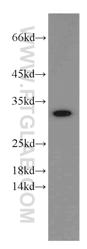
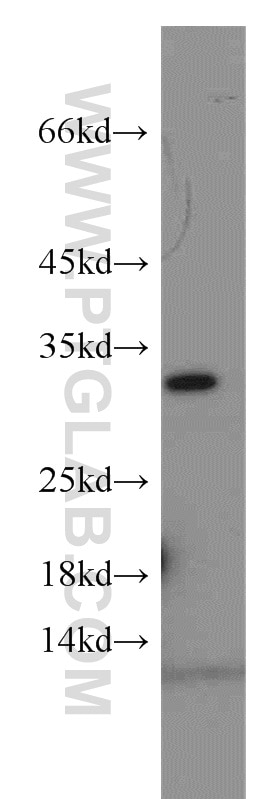
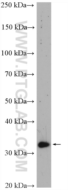
| Positive WB detected in | HepG2 cells, mouse brain tissue, HuH-7 cells |
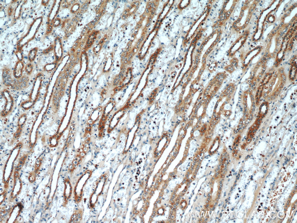
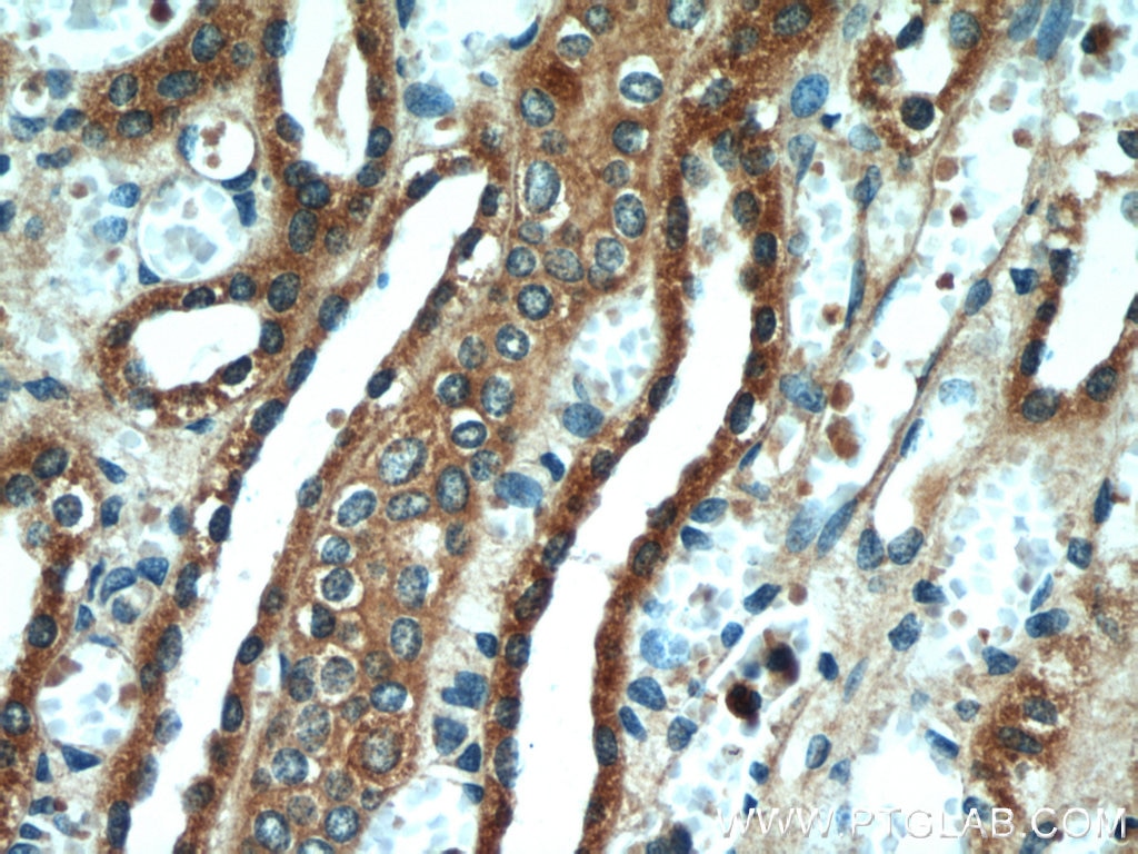
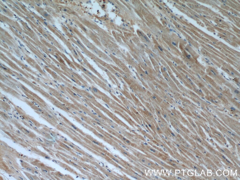
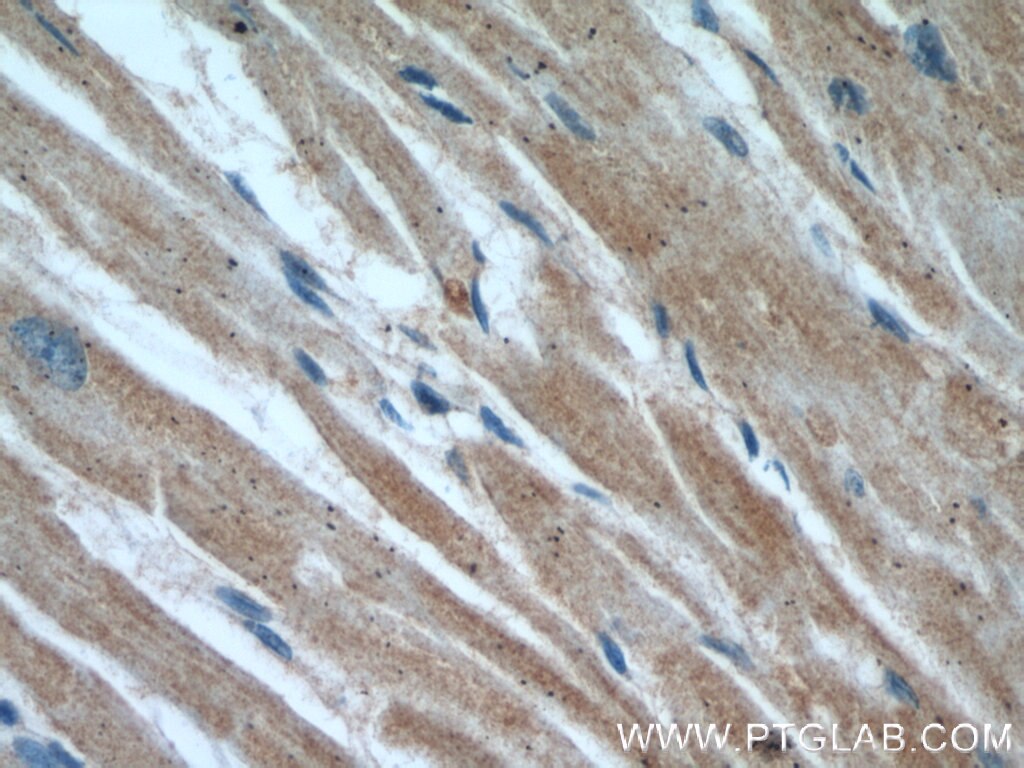
| Positive IHC detected in | human kidney tissue, human heart tissue Note: suggested antigen retrieval with TE buffer pH 9.0; (*) Alternatively, antigen retrieval may be performed with citrate buffer pH 6.0 |
Recommended dilution
| Application | Dilution |
|---|---|
| Western Blot (WB) | WB : 1:500-1:1000 |
| Immunohistochemistry (IHC) | IHC : 1:20-1:200 |
| It is recommended that this reagent should be titrated in each testing system to obtain optimal results. | |
| Sample-dependent, Check data in validation data gallery. | |
Published Applications
| WB | See 3 publications below |
Product Information
55261-1-AP targets VDAC2 in WB, IHC, ELISA applications and shows reactivity with human, mouse, rat samples.
| Tested Reactivity | human, mouse, rat |
| Cited Reactivity | human, mouse |
| Host / Isotype | Rabbit / IgG |
| Class | Polyclonal |
| Type | Antibody |
| Immunogen |
Peptide 相同性解析による交差性が予測される生物種 |
| Full Name | voltage-dependent anion channel 2 |
| Calculated molecular weight | 33 kDa |
| Observed molecular weight | 31-33 kDa |
| GenBank accession number | NM_003375 |
| Gene Symbol | VDAC2 |
| Gene ID (NCBI) | 7417 |
| RRID | AB_10852961 |
| Conjugate | Unconjugated |
| Form | |
| Form | Liquid |
| Purification Method | Antigen affinity purification |
| UNIPROT ID | P45880 |
| Storage Buffer | PBS with 0.02% sodium azide and 50% glycerol{{ptg:BufferTemp}}7.3 |
| Storage Conditions | Store at -20°C. Aliquoting is unnecessary for -20oC storage. |
Background Information
VDAC2 belongs to the eukaryotic mitochondrial porin family. It forms a channel through the mitochondrial outer membrane that allows diffusion of small hydrophilic molecules. This antibody is specific to VDAC2.
Protocols
| Product Specific Protocols | |
|---|---|
| IHC protocol for VDAC2 antibody 55261-1-AP | Download protocol |
| WB protocol for VDAC2 antibody 55261-1-AP | Download protocol |
| Standard Protocols | |
|---|---|
| Click here to view our Standard Protocols |
Publications
| Species | Application | Title |
|---|---|---|
Cell Physiol Biochem Knockout of VDAC1 in H9c2 Cells Promotes Oxidative Stress-Induced Cell Apoptosis through Decreased Mitochondrial Hexokinase II Binding and Enhanced Glycolytic Stress. | ||
J Mol Med (Berl) Cerium oxide nanoparticles as potent inhibitors of ferroptosis: role of antioxidant activity and protein regulation |

