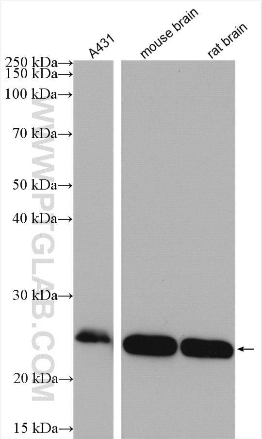
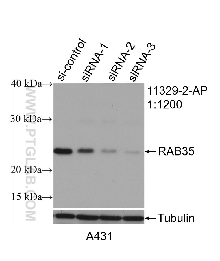
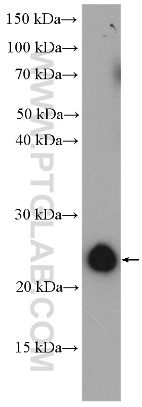
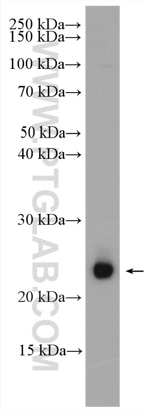
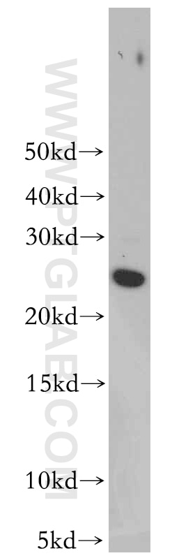
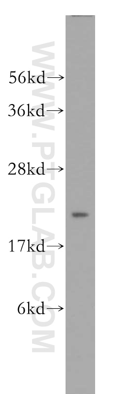
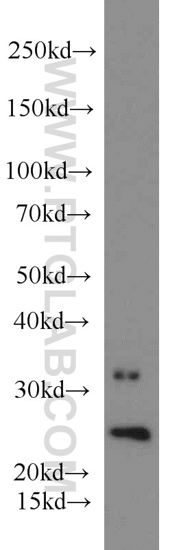
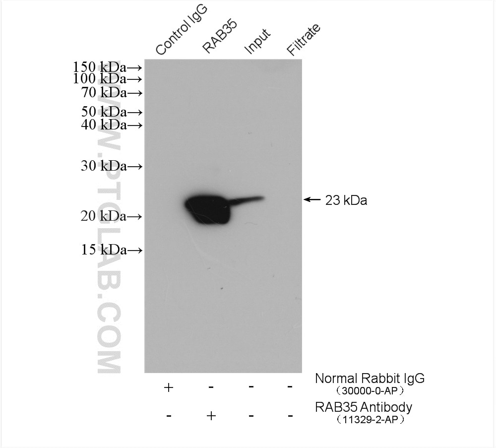
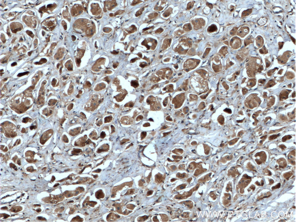
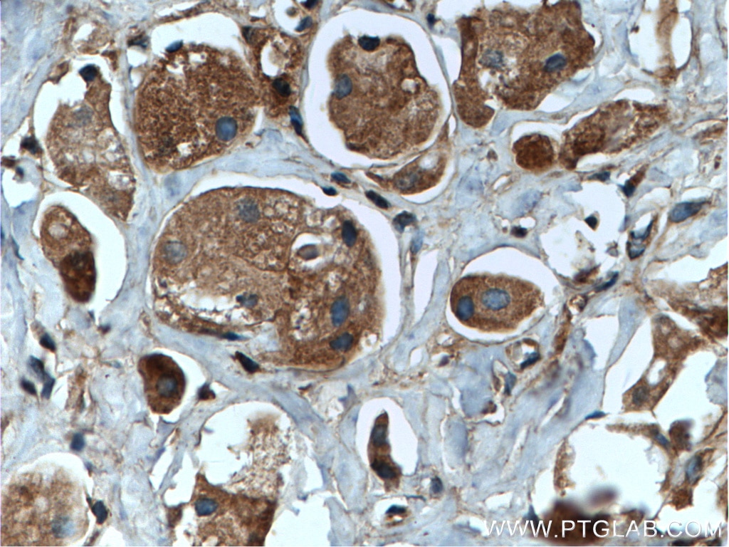
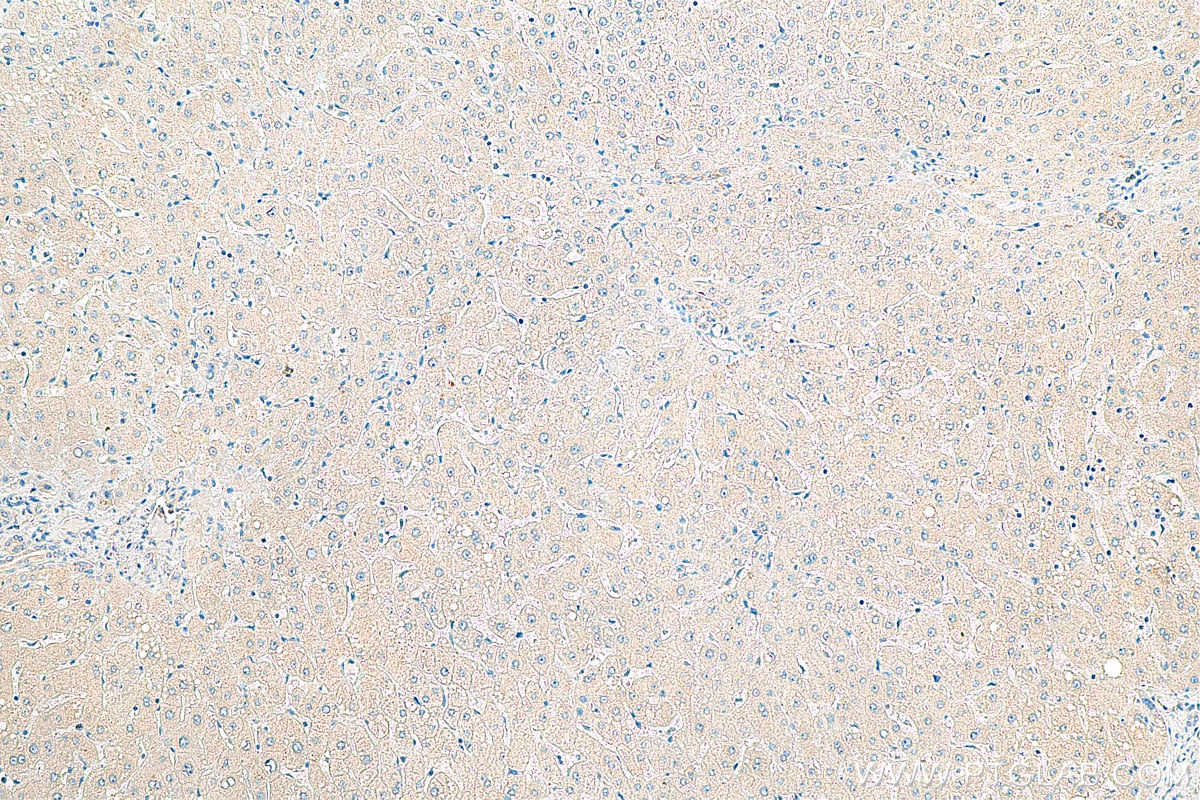
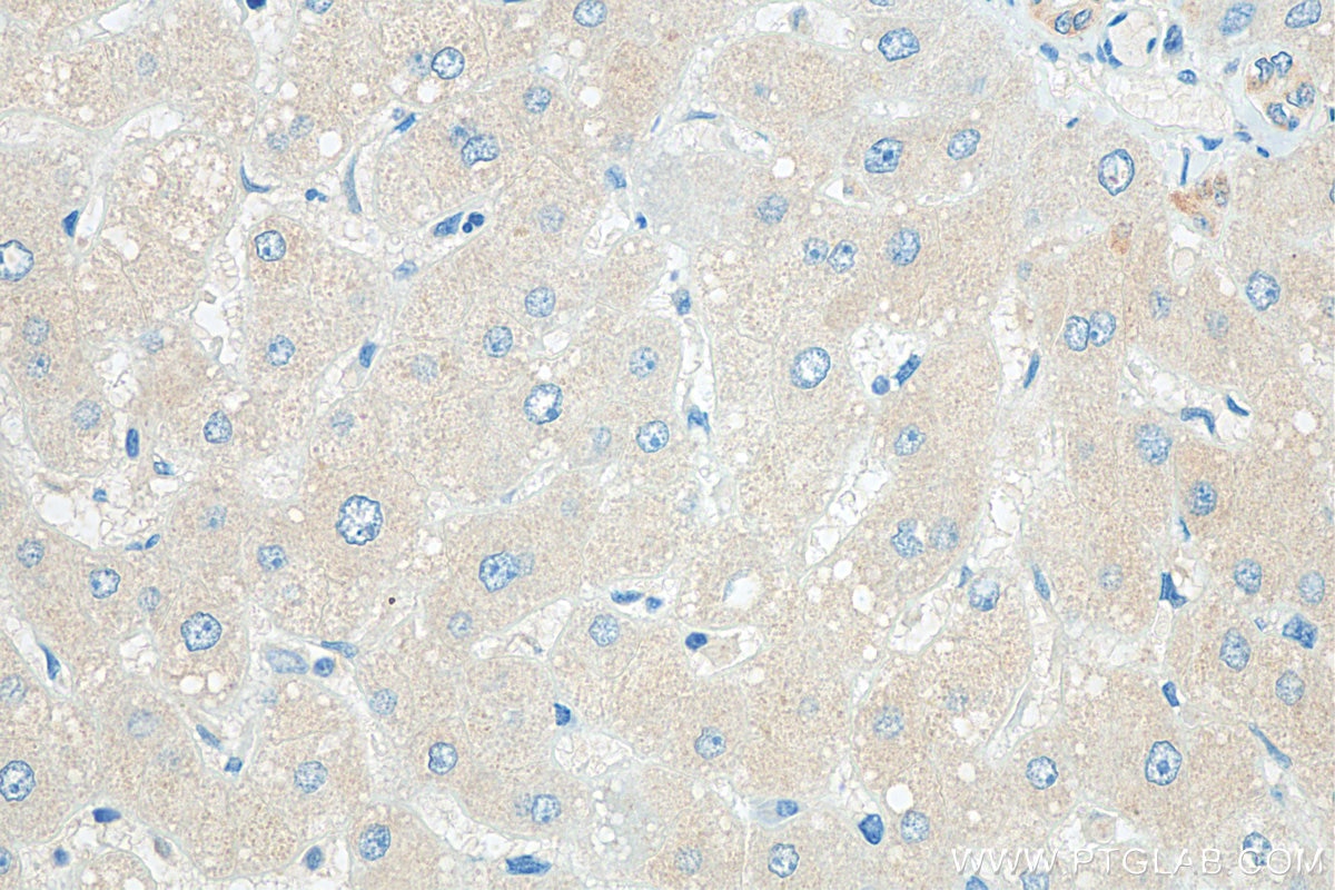
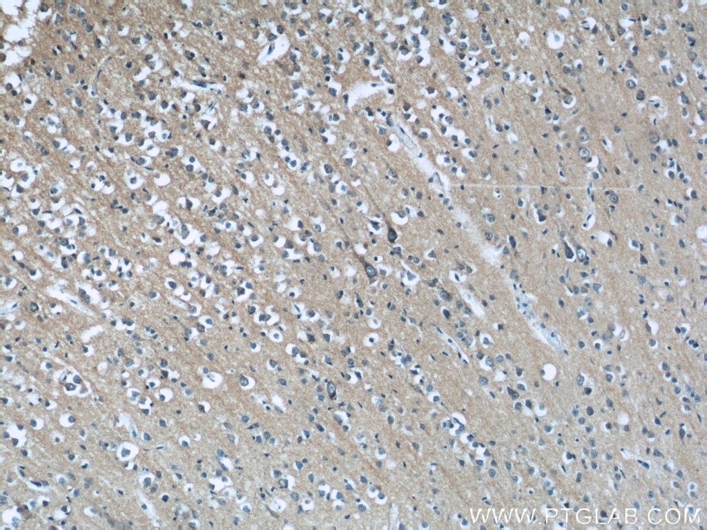
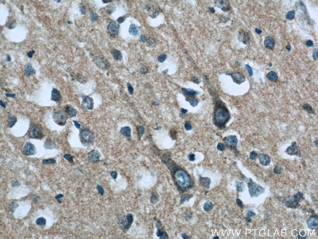
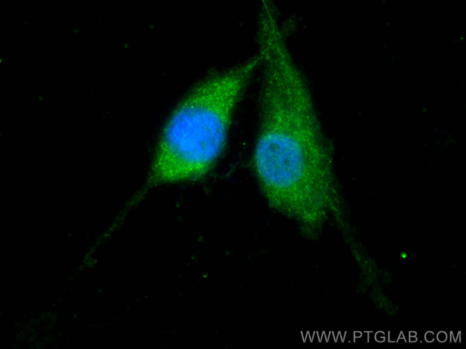
Validation Data Gallery
Tested Applications
| Positive WB detected in | A431 cells, A375 cells, human brain tissue, HeLa cells, MCF-7 cells, RAW 264.7 cells, mouse brain tissue, rat brain tissue |
| Positive IP detected in | mouse brain tissue |
| Positive IHC detected in | human breast cancer tissue, human liver tissue, human brain tissue Note: suggested antigen retrieval with TE buffer pH 9.0; (*) Alternatively, antigen retrieval may be performed with citrate buffer pH 6.0 |
| Positive IF/ICC detected in | U-87 MG cells |
Recommended dilution
| Application | Dilution |
|---|---|
| Western Blot (WB) | WB : 1:1000-1:6000 |
| Immunoprecipitation (IP) | IP : 0.5-4.0 ug for 1.0-3.0 mg of total protein lysate |
| Immunohistochemistry (IHC) | IHC : 1:50-1:500 |
| Immunofluorescence (IF)/ICC | IF/ICC : 1:200-1:800 |
| It is recommended that this reagent should be titrated in each testing system to obtain optimal results. | |
| Sample-dependent, Check data in validation data gallery. | |
Published Applications
| KD/KO | See 12 publications below |
| WB | See 39 publications below |
| IHC | See 2 publications below |
| IF | See 18 publications below |
| IP | See 1 publications below |
| CoIP | See 1 publications below |
Product Information
11329-2-AP targets RAB35 in WB, IHC, IF/ICC, IP, CoIP, ELISA applications and shows reactivity with human, mouse, rat samples.
| Tested Reactivity | human, mouse, rat |
| Cited Reactivity | human, mouse, rat, pig, canine |
| Host / Isotype | Rabbit / IgG |
| Class | Polyclonal |
| Type | Antibody |
| Immunogen |
CatNo: Ag1872 Product name: Recombinant human RAB35 protein Source: e coli.-derived, PGEX-4T Tag: GST Domain: 1-201 aa of BC015931 Sequence: MARDYDHLFKLLIIGDSGVGKSSLLLRFADNTFSGSYITTIGVDFKIRTVEINGEKVKLQIWDTAGQERFRTITSTYYRGTHGVIVVYDVTSAESFVNVKRWLHEINQNCDDVCRILVGNKNDDPERKVVETEDAYKFAGQMGIQLFETSAKENVNVEEMFNCITELVLRAKKDNLAKQQQQQQNDVVKLTKNSKRKKRCC 相同性解析による交差性が予測される生物種 |
| Full Name | RAB35, member RAS oncogene family |
| Calculated molecular weight | 201 aa, 23 kDa |
| Observed molecular weight | 23 kDa |
| GenBank accession number | BC015931 |
| Gene Symbol | RAB35 |
| Gene ID (NCBI) | 11021 |
| RRID | AB_2238179 |
| Conjugate | Unconjugated |
| Form | |
| Form | Liquid |
| Purification Method | Antigen affinity purification |
| UNIPROT ID | Q15286 |
| Storage Buffer | PBS with 0.02% sodium azide and 50% glycerol{{ptg:BufferTemp}}7.3 |
| Storage Conditions | Store at -20°C. Stable for one year after shipment. Aliquoting is unnecessary for -20oC storage. |
Background Information
RAB35 is a member of the small GTPase superfamily. Like the other Rab proteins, RAB35 has 2 consecutive cysteine residues at its C terminus, and this region is thought to be involved in membrane association. Rab35 is found on the cell surface and endosomes, where it has been proposed to play a role in recycling of the transferrin and other receptors.
Protocols
| Product Specific Protocols | |
|---|---|
| IF protocol for RAB35 antibody 11329-2-AP | Download protocol |
| IHC protocol for RAB35 antibody 11329-2-AP | Download protocol |
| IP protocol for RAB35 antibody 11329-2-AP | Download protocol |
| WB protocol for RAB35 antibody 11329-2-AP | Download protocol |
| Standard Protocols | |
|---|---|
| Click here to view our Standard Protocols |
Publications
| Species | Application | Title |
|---|---|---|
Cell Metab Oligodendrocytes Provide Antioxidant Defense Function for Neurons by Secreting Ferritin Heavy Chain. | ||
Nat Cell Biol Protein kinase N controls a lysosomal lipid switch to facilitate nutrient signalling via mTORC1. | ||
Nat Commun A RAB35-p85/PI3K axis controls oscillatory apical protrusions required for efficient chemotactic migration.
|

