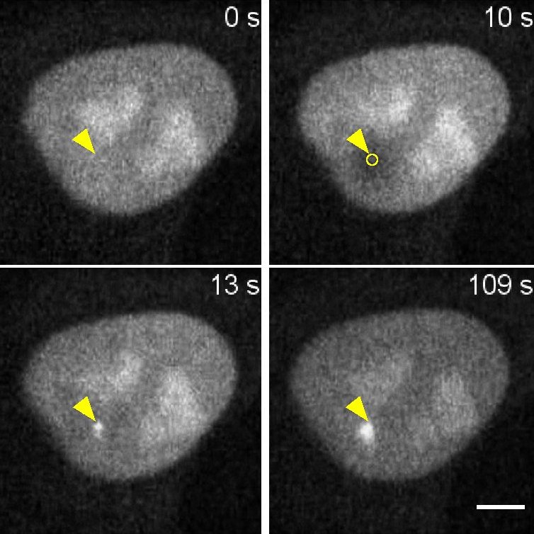
Validation Data Gallery
Product Information
DNA plasmid encoding for anti-PARP1 VHH (anti-PARP1 Nanobody) fused to TagRFP.
| Description | The PARP1-Chromobody visualizes endogenous PARP1 in real time in live cells.
• Easily transfect your cells • Use PARP1-Chromobody® as marker of DNA damage after microirradiation • PARP1-Chromobody® does not affect cell viability |
| Applications | Live cell imaging |
| Specificity | Poly(ADP-ribose) polymerase 1 (PARP1), tested in human cells. Does not bind to PARP2, PARP3, andPARP 9 |
| Reporter | TagRFP |
| Vector type | Mammalian expression vector |
| Promoter | Constitutive CMV IE |
| Codon usage | Mammalian |
| Selection | Kan/Neo |
| Sequence | With the PARP1-Chromobody plasmid you receive the sequence information of the alpaca antibody to PARP1 fused to TagGFP2 or TagRFP, as well as the full vector sequence. |
| Transfection | Transfection of Chromobody plasmids into mammalian cells can be done with standard DNA-transfection methods, e.g. lipofection (Lipofectamine 2000® from Thermo Fisher Scientific), according to the manufacturer’s protocol for the transfection reagent. Please choose the transfection method that works the best for your cell type. |
| Microscopy techniques | Wide-field epifluorescence microscopy; confocal microscopy |
| Storage Condition | Shipped at ambient temperature. Store at -20 °C |
Documentation
| SDS |
|---|
| xcr_SDS_PARP1-Chromobody® plasmid (TagRFP) (EN) |
| Datasheet |
|---|
| PARP1-Chromobody® plasmid (TagRFP) Datasheet |
| Brochure |
|---|
| Chromotek nanobodies brochure (PDF) |
Publications
| Application | Title |
|---|---|
Proc Natl Acad Sci U S A Rapid recruitment of p53 to DNA damage sites directs DNA repair choice and integrity. | |
J Cell Sci Local inhibition of rRNA transcription without nucleolar segregation after targeted ion irradiation of the nucleolus. | |
J Biophotonics Chromatin nanoscale compaction in live cells visualized by acceptor-to-donor ratio corrected Förster resonance energy transfer between DNA dyes. | |
Biophys J Measuring PARP1 mobility at DNA damage sites by Segmented Fluorescence Correlation Spectroscopy (FCS) | |
Sci Rep Segmented fluorescence correlation spectroscopy (FCS) on a commercial laser scanning microscope |


