
Validation Data Gallery
Tested Applications
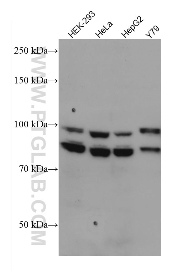
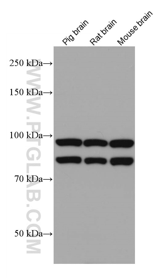
| Positive WB detected in | HEK-293 cells, pig brain tissue, HeLa cells, HepG2 cells, Y79 cells, mouse brain tissue, rat brain tissue |
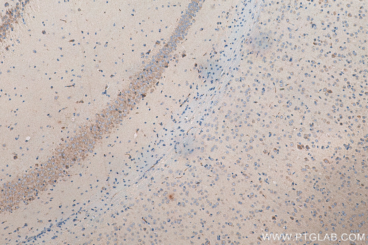
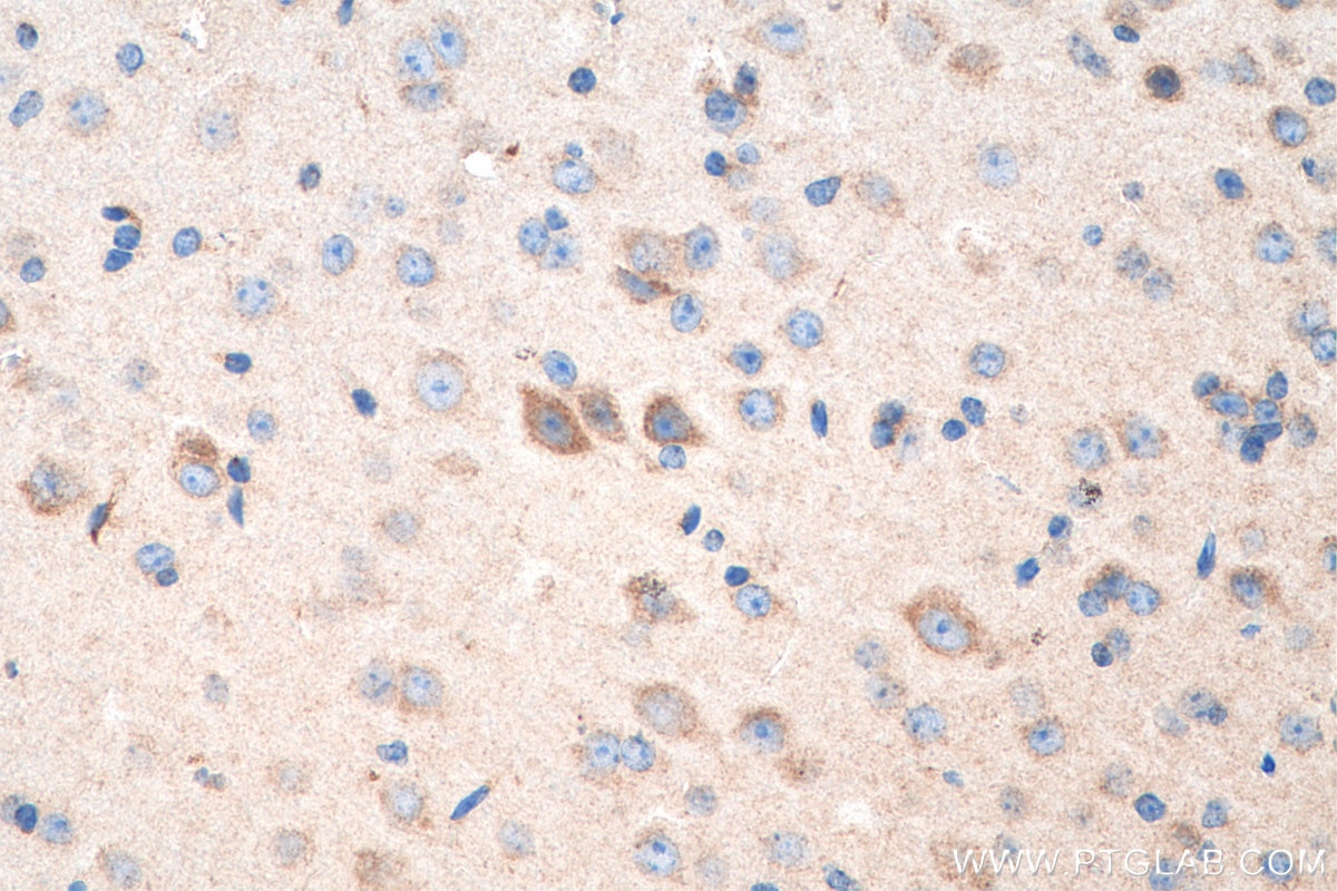
| Positive IHC detected in | mouse brain tissue Note: suggested antigen retrieval with TE buffer pH 9.0; (*) Alternatively, antigen retrieval may be performed with citrate buffer pH 6.0 |
Recommended dilution
| Application | Dilution |
|---|---|
| Western Blot (WB) | WB : 1:500-1:2000 |
| Immunohistochemistry (IHC) | IHC : 1:400-1:1600 |
| It is recommended that this reagent should be titrated in each testing system to obtain optimal results. | |
| Sample-dependent, Check data in validation data gallery. | |
Published Applications
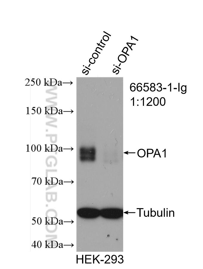
| KD/KO | See 1 publications below |
| WB | See 27 publications below |
| IF | See 4 publications below |
Product Information
66583-1-Ig targets OPA1 in WB, IHC, IF, ELISA applications and shows reactivity with Human, mouse, pig, rat samples.
| Tested Reactivity | Human, mouse, pig, rat |
| Cited Reactivity | human, mouse, rat, fish |
| Host / Isotype | Mouse / IgG2b |
| Class | Monoclonal |
| Type | Antibody |
| Immunogen |
CatNo: Ag26868 Product name: Recombinant human OPA1 protein Source: e coli.-derived, PGEX-4T Tag: GST Domain: 650-780 aa of BC075805 Sequence: NTTVDIKLKQWTDKQLPNKAVEVAWETLQEEFSRFMTEPKGKEHDDIFDKLKEAVKEESIKRHKWNDFAEDSLRVIQHNALEDRSISDKQQWDAAIYFMEEALQARLKDTENAIENMVGPDWKKRWLYWKN 相同性解析による交差性が予測される生物種 |
| Full Name | optic atrophy 1 (autosomal dominant) |
| Calculated molecular weight | 960 aa, 112 kDa |
| Observed molecular weight | 100 kDa and 80-90 kDa |
| GenBank accession number | BC075805 |
| Gene Symbol | OPA1 |
| Gene ID (NCBI) | 4976 |
| RRID | AB_2881943 |
| Conjugate | Unconjugated |
| Form | |
| Form | Liquid |
| Purification Method | Protein A purification |
| UNIPROT ID | O60313 |
| Storage Buffer | PBS with 0.02% sodium azide and 50% glycerol{{ptg:BufferTemp}}7.3 |
| Storage Conditions | Store at -20°C. Stable for one year after shipment. Aliquoting is unnecessary for -20oC storage. |
Background Information
OPA1 is a nuclear-encoded mitochondrial protein with similarity to dynamin-related GTPases. OPA1 localizes to the inner mitochondrial membrane and helps regulate mitochondrial stability and energy output. This protein also sequesters cytochrome c. OPA1 is associated with the inner membrane and protects cells from apoptosis by regulating inner membrane dynamics. Mutation of OPA1 causes the disease dominant optic atrophy, a degeneration of the retinal ganglion cells. OPA1 undergoes complex posttranscriptional regulation and posttranslational proteolysis. OPA1 is regulated by proteolytic cleavage, which degrades long OPA1 isoforms into short isoforms. The gene OPA1 can be cleaved into some chains with MW 100 kDa and 80-90 kDa.
Protocols
| Product Specific Protocols | |
|---|---|
| IHC protocol for OPA1 antibody 66583-1-Ig | Download protocol |
| WB protocol for OPA1 antibody 66583-1-Ig | Download protocol |
| Standard Protocols | |
|---|---|
| Click here to view our Standard Protocols |
Publications
| Species | Application | Title |
|---|---|---|
J Nanobiotechnology Human periodontal ligament stem cell sheets activated by graphene oxide quantum dots repair periodontal bone defects by promoting mitochondrial dynamics dependent osteogenic differentiation | ||
Genes Dis TNFα-reliant FSP1 up-regulation promotes intervertebral disc degeneration via caspase 3-dependent apoptosis | ||
Adv Sci (Weinh) HIGD1A Alleviates Oxidative Stress Related Ovarian Hypofunction by Enhancing Granulosa Cell Functions via NF-κB/SOD2 Signaling Pathway | ||
Adv Sci (Weinh) Ubiquitination of ACSL4 by Parkin Suppresses Ferroptosis and Rescues Glucocorticoid-Induced Bone Loss | ||
Cell Death Dis Cadmium-induced apoptosis of Leydig cells is mediated by excessive mitochondrial fission and inhibition of mitophagy | ||