WB Figures
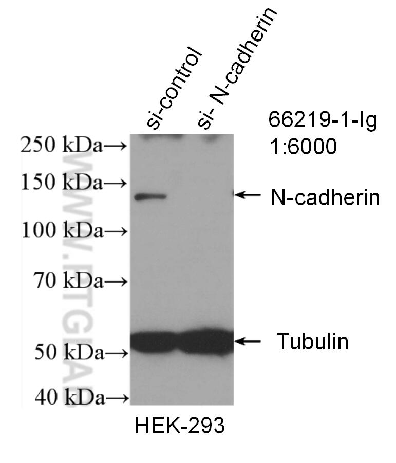
WB analysis of HEK-293 using 66219-1-Ig (same clone as 66219-1-PBS)
WB result of N-cadherin antibody (66219-1-Ig; 1:6000; incubated at room temperature for 1.5 hours) with sh-Control and sh-N-cadherin transfected HEK-293 cells. This data was developed using the same antibody clone with 66219-1-PBS in a different storage buffer formulation.
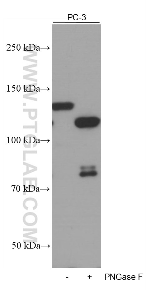
WB analysis of PC-3 using 66219-1-Ig (same clone as 66219-1-PBS)
Untreated and PNGase F-treated lysates of PC-3 cells were subjected to SDS PAGE followed by western blot with 66219-1-Ig (N-cadherin antibody) at dilution of 1:5000 incubated at room temperature for 1.5 hours. PNGase F was obtained from Atagenix (cat.NO. ata808). This data was developed using the same antibody clone with 66219-1-PBS in a different storage buffer formulation.
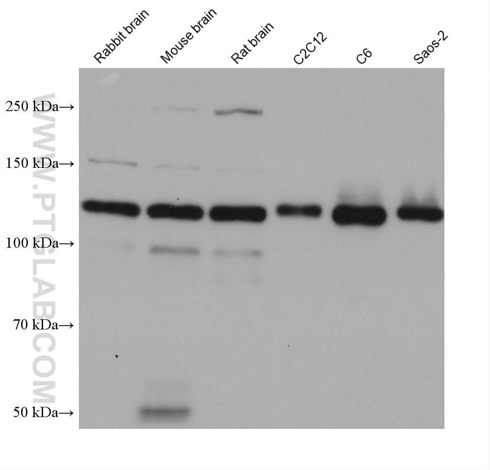
WB analysis using 66219-1-Ig (same clone as 66219-1-PBS)
Various lysates were subjected to SDS PAGE followed by western blot with 66219-1-Ig (N-cadherin antibody) at dilution of 1:20000 incubated at room temperature for 1.5 hours. This data was developed using the same antibody clone with 66219-1-PBS in a different storage buffer formulation.
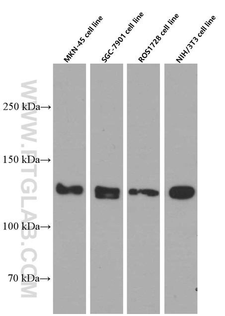
WB analysis of MKN-45 using 66219-1-Ig (same clone as 66219-1-PBS)
Various lysates were subjected to SDS PAGE followed by western blot with 66219-1-Ig (N-cadherin Antibody) at dilution of 1:4000 incubated at room temperature for 1.5 hours. This data was developed using the same antibody clone with 66219-1-PBS in a different storage buffer formulation.
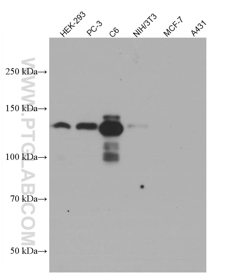
WB analysis using 66219-1-Ig (same clone as 66219-1-PBS)
HEK-293 (N-cadherin+), PC-3 cells (N-cadherin+), C6 cells (N-cadherin+), NIH/3T3 cells (N-cadherin+), MCF-7 (N-cadherin-), A431 (N-cadherin-) cell lysates were subjected to SDS PAGE followed by western blot with 66219-1-Ig (N-cadherin antibody) at dilution of 1:5000 incubated at room temperature for 1.5 hours. This data was developed using the same antibody clone with 66219-1-PBS in a different storage buffer formulation.
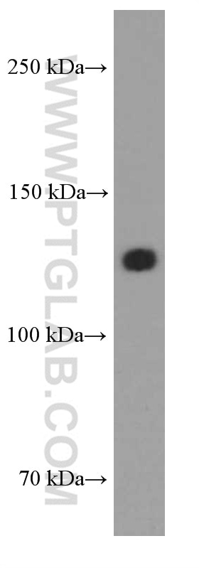
WB analysis of human heart using 66219-1-Ig (same clone as 66219-1-PBS)
human heart tissue were subjected to SDS PAGE followed by western blot with 66219-1-Ig (N-cadherin antibody) at dilution of 1:2000 incubated at room temperature for 1.5 hours. This data was developed using the same antibody clone with 66219-1-PBS in a different storage buffer formulation.
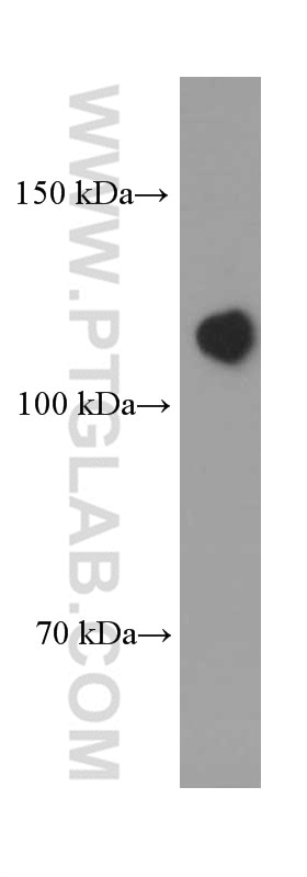
WB analysis of pig brain using 66219-1-Ig (same clone as 66219-1-PBS)
pig brain tissue were subjected to SDS PAGE followed by western blot with 66219-1-Ig (N-cadherin Antibody) at dilution of 1:2000 incubated at room temperature for 1.5 hours. This data was developed using the same antibody clone with 66219-1-PBS in a different storage buffer formulation.
IHC Figures
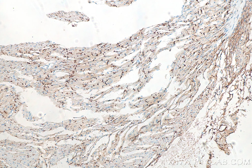
IHC staining of mouse heart using 66219-1-Ig (same clone as 66219-1-PBS)
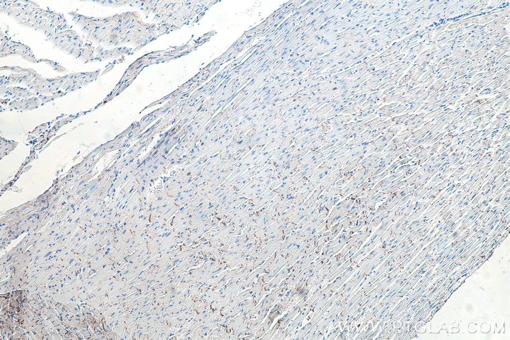
Immunohistochemical analysis of paraffin-embedded mouse heart tissue slide using 66219-1-Ig (N-cadherin antibody) at dilution of 1:15000 (under 10x lens). Heat mediated antigen retrieval with Tris-EDTA buffer (pH 9.0). This data was developed using the same antibody clone with 66219-1-PBS in a different storage buffer formulation.
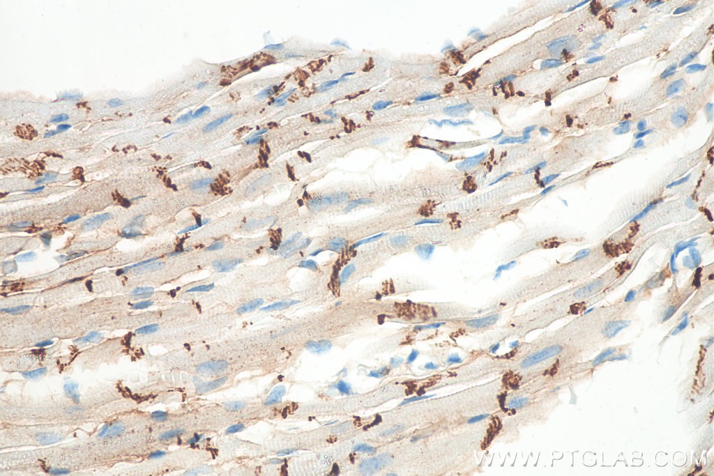
IHC staining of mouse heart using 66219-1-Ig (same clone as 66219-1-PBS)
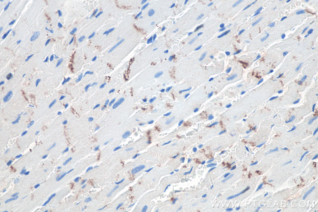
Immunohistochemical analysis of paraffin-embedded mouse heart tissue slide using 66219-1-Ig (N-cadherin antibody) at dilution of 1:15000 (under 40x lens). Heat mediated antigen retrieval with Tris-EDTA buffer (pH 9.0). This data was developed using the same antibody clone with 66219-1-PBS in a different storage buffer formulation.
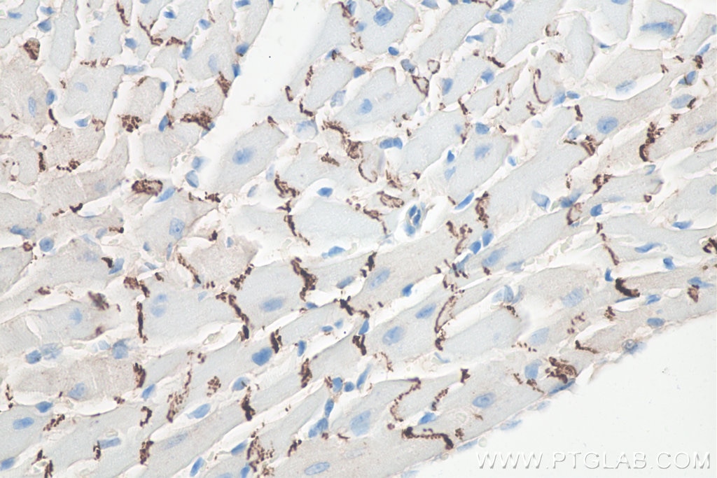
IHC staining of mouse heart using 66219-1-Ig (same clone as 66219-1-PBS)
Immunohistochemical analysis of paraffin-embedded mouse heart tissue slide using 66219-1-Ig (N-cadherin antibody) at dilution of 1:6000 (under 40x lens). Heat mediated antigen retrieval with Tris-EDTA buffer (pH 9.0). This data was developed using the same antibody clone with 66219-1-PBS in a different storage buffer formulation.
IHC staining of rat heart using 66219-1-Ig (same clone as 66219-1-PBS)
Immunohistochemical analysis of paraffin-embedded rat heart tissue slide using 66219-1-Ig (N-cadherin antibody) at dilution of 1:6000 (under 40x lens). Heat mediated antigen retrieval with Tris-EDTA buffer (pH 9.0). This data was developed using the same antibody clone with 66219-1-PBS in a different storage buffer formulation.
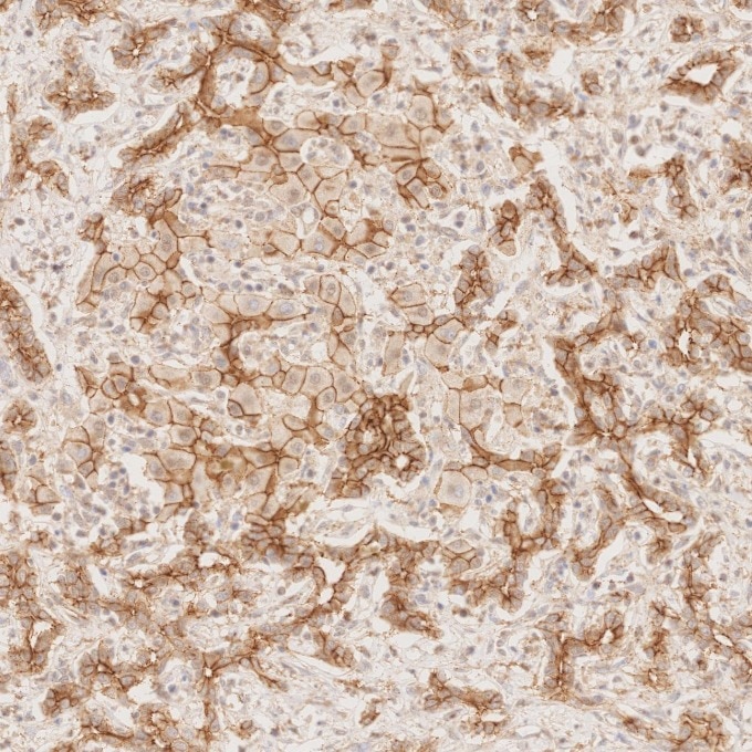
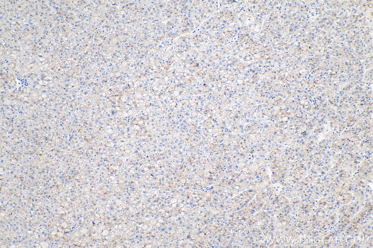
IHC staining of human liver cancer using 66219-1-Ig (same clone as 66219-1-PBS)
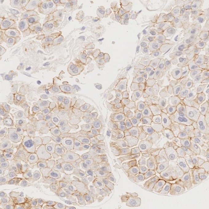
Immunohistochemical analysis of paraffin-embedded human liver cancer tissue slide using 66219-1-Ig (N-cadherin antibody) at dilution of 1:6000 (under 10x lens). Heat mediated antigen retrieval with Tris-EDTA buffer (pH 9.0). This data was developed using the same antibody clone with 66219-1-PBS in a different storage buffer formulation.
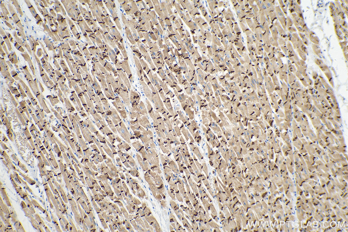
IHC staining of human heart using 66219-1-Ig (same clone as 66219-1-PBS)
Immunohistochemical analysis of paraffin-embedded human heart tissue slide using 66219-1-Ig (N-cadherin antibody) at dilution of 1:6000 (under 10x lens). Heat mediated antigen retrieval with Tris-EDTA buffer (pH 9.0). This data was developed using the same antibody clone with 66219-1-PBS in a different storage buffer formulation.
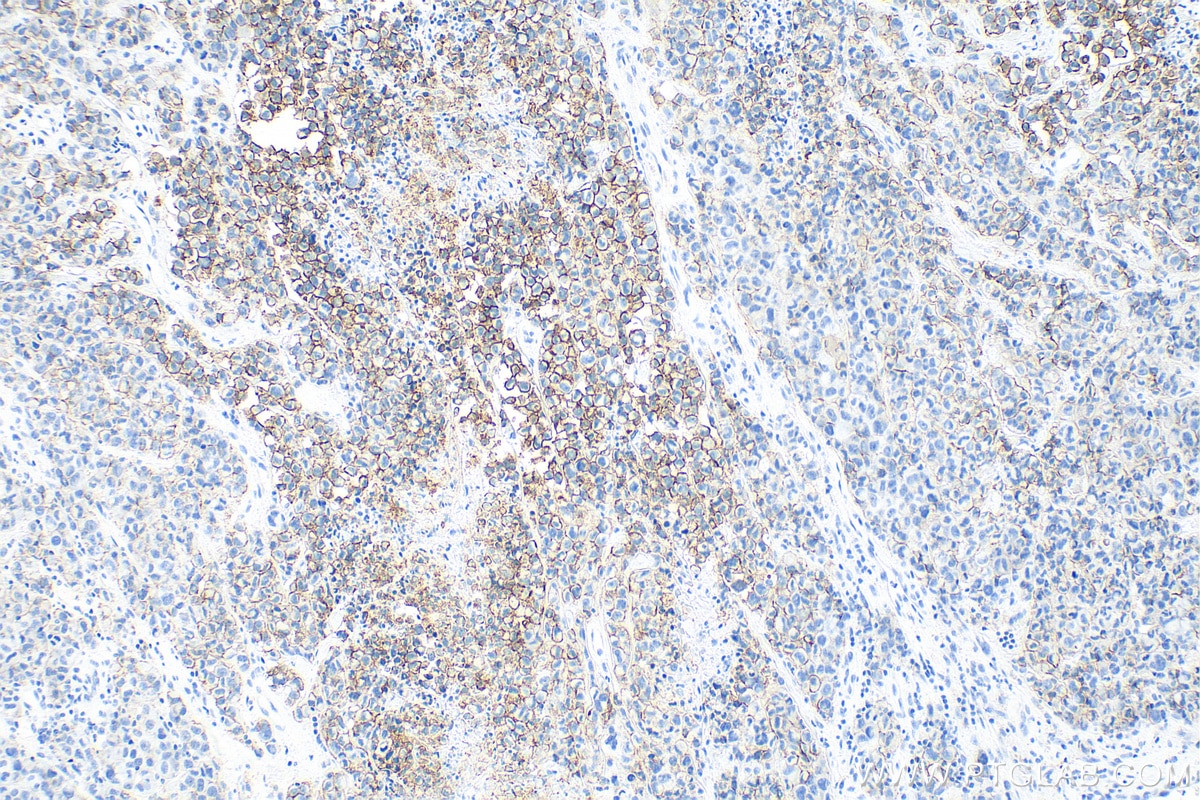
IHC staining of human malignant melanoma using 66219-1-Ig (same clone as 66219-1-PBS)
Immunohistochemical analysis of paraffin-embedded human malignant melanoma tissue slide using 66219-1-Ig (N-cadherin antibody) at dilution of 1:5000 (under 10x lens). Heat mediated antigen retrieval with Tris-EDTA buffer (pH 9.0). This data was developed using the same antibody clone with 66219-1-PBS in a different storage buffer formulation.
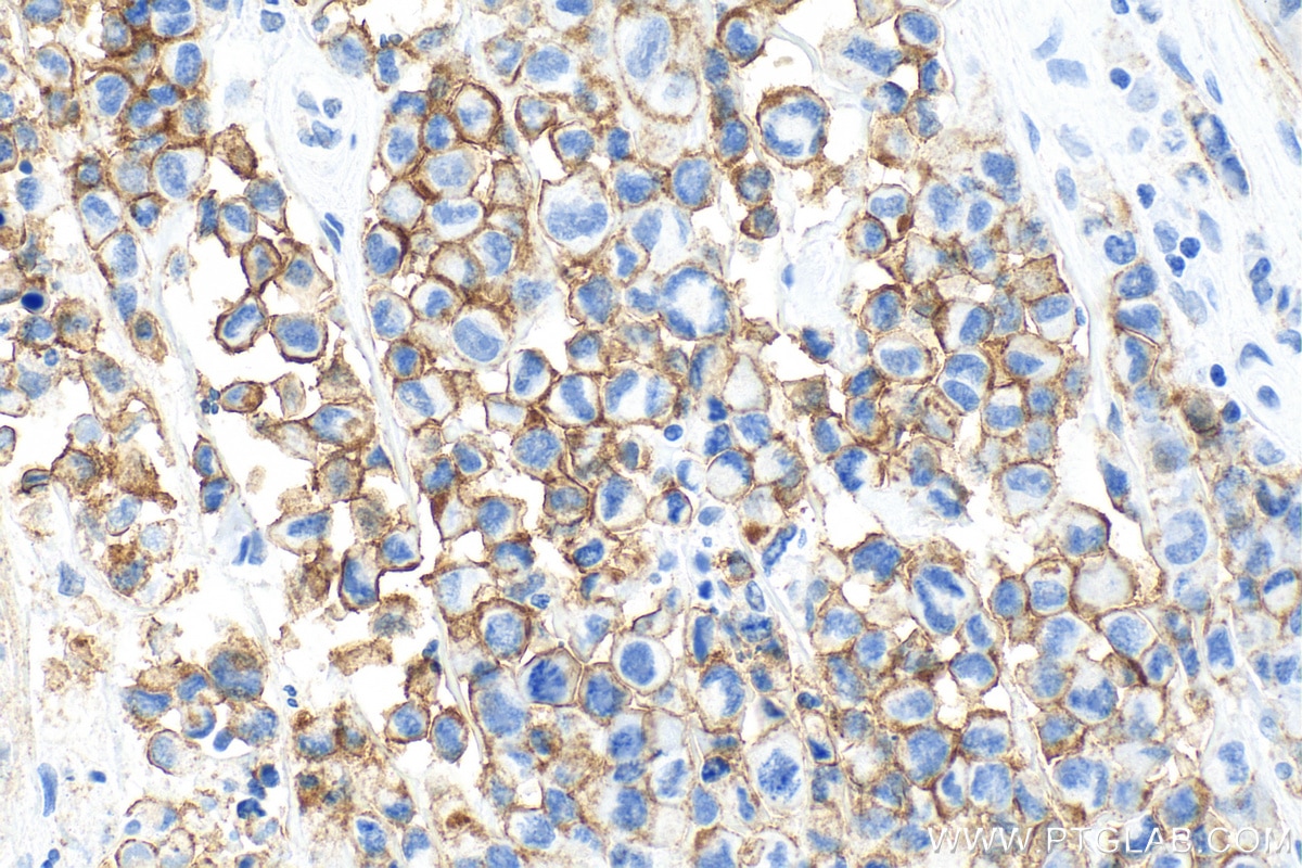
IHC staining of human malignant melanoma using 66219-1-Ig (same clone as 66219-1-PBS)
Immunohistochemical analysis of paraffin-embedded human malignant melanoma tissue slide using 66219-1-Ig (N-cadherin antibody) at dilution of 1:5000 (under 40x lens). Heat mediated antigen retrieval with Tris-EDTA buffer (pH 9.0). This data was developed using the same antibody clone with 66219-1-PBS in a different storage buffer formulation.
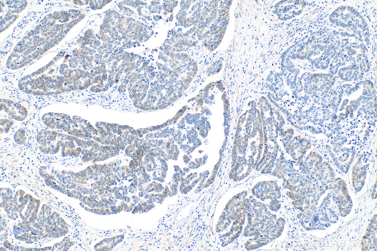
IHC staining of human ovary tumor using 66219-1-Ig (same clone as 66219-1-PBS)
Immunohistochemical analysis of paraffin-embedded human ovary tumor tissue slide using 66219-1-Ig (N-cadherin antibody) at dilution of 1:5000 (under 10x lens). Heat mediated antigen retrieval with Tris-EDTA buffer (pH 9.0). This data was developed using the same antibody clone with 66219-1-PBS in a different storage buffer formulation.
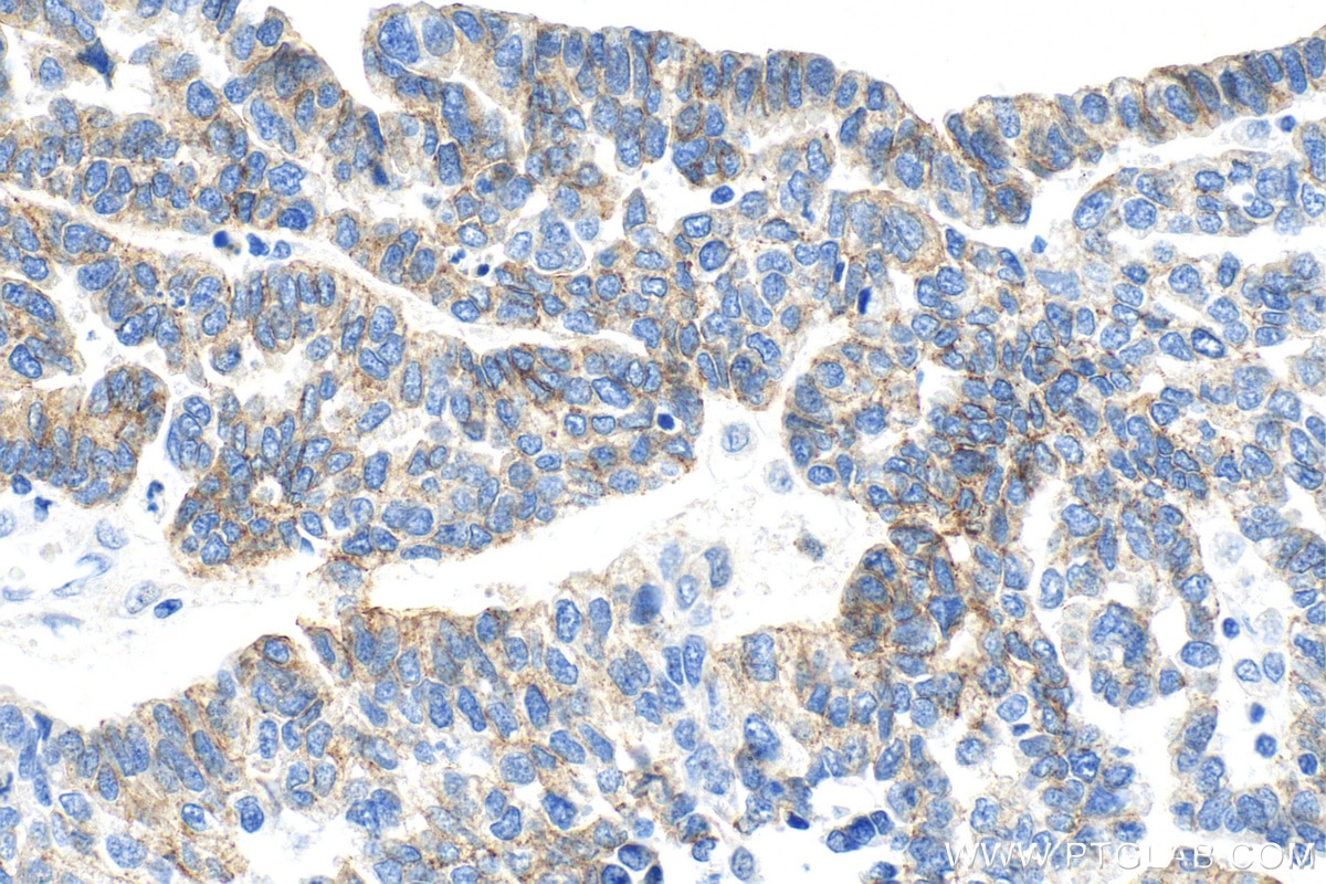
IHC staining of human ovary tumor using 66219-1-Ig (same clone as 66219-1-PBS)
Immunohistochemical analysis of paraffin-embedded human ovary tumor tissue slide using 66219-1-Ig (N-cadherin antibody) at dilution of 1:5000 (under 40x lens). Heat mediated antigen retrieval with Tris-EDTA buffer (pH 9.0). This data was developed using the same antibody clone with 66219-1-PBS in a different storage buffer formulation.
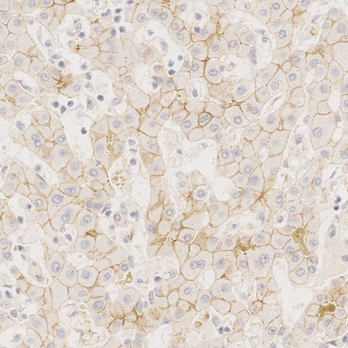
IHC staining of human liver using 66219-1-Ig (same clone as 66219-1-PBS)
Immunohistochemical analysis of paraffin-embedded human liver tissue slide using 66219-1-Ig (N-cadherin antibody) at dilution of 1:6000. Heat mediated antigen retrieval with Tris-EDTA buffer (pH 9.0). This data was developed using the same antibody clone with 66219-1-PBS in a different storage buffer formulation.
IHC staining of Cholangiocarcinoma using 66219-1-Ig (same clone as 66219-1-PBS)
Immunohistochemical analysis of paraffin-embedded Cholangiocarcinoma tissue slide using 66219-1-Ig (N-cadherin antibody) at dilution of 1:6000. Heat mediated antigen retrieval with Tris-EDTA buffer (pH 9.0). This data was developed using the same antibody clone with 66219-1-PBS in a different storage buffer formulation.
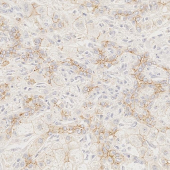
IHC staining of human liver cancer using 66219-1-Ig (same clone as 66219-1-PBS)
Immunohistochemical analysis of paraffin-embedded human liver cancer tissue slide using 66219-1-Ig (N-cadherin antibody) at dilution of 1:6000. Heat mediated antigen retrieval with Tris-EDTA buffer (pH 9.0). This data was developed using the same antibody clone with 66219-1-PBS in a different storage buffer formulation.
IHC staining of human heart using 66219-1-Ig (same clone as 66219-1-PBS)
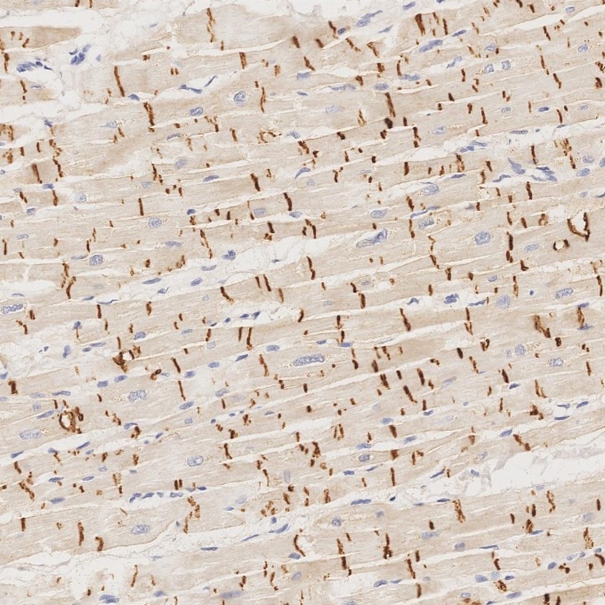
Immunohistochemical analysis of paraffin-embedded human heart tissue slide using 66219-1-Ig (N-cadherin antibody) at dilution of 1:6000. Heat mediated antigen retrieval with Tris-EDTA buffer (pH 9.0). This data was developed using the same antibody clone with 66219-1-PBS in a different storage buffer formulation.
IHC staining of rat heart using 66219-1-Ig (same clone as 66219-1-PBS)
Immunohistochemical analysis of paraffin-embedded rat heart tissue slide using 66219-1-Ig (N-cadherin antibody) at dilution of 1:8000 (under 10x lens). Heat mediated antigen retrieval with Tris-EDTA buffer (pH 9.0). This data was developed using the same antibody clone with 66219-1-PBS in a different storage buffer formulation.
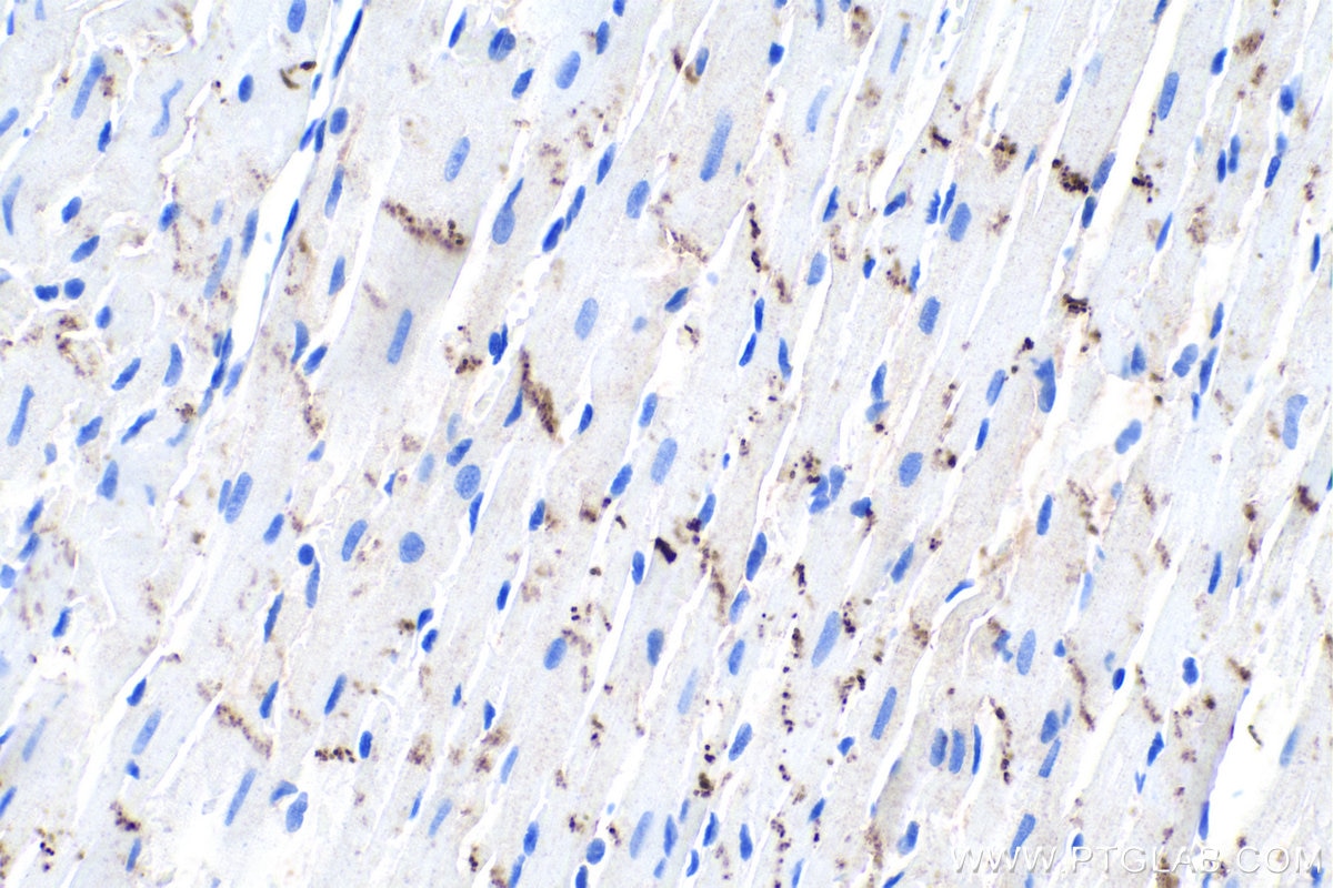
IHC staining of rat heart using 66219-1-Ig (same clone as 66219-1-PBS)
Immunohistochemical analysis of paraffin-embedded rat heart tissue slide using 66219-1-Ig (N-cadherin antibody) at dilution of 1:8000 (under 40x lens). Heat mediated antigen retrieval with Tris-EDTA buffer (pH 9.0). This data was developed using the same antibody clone with 66219-1-PBS in a different storage buffer formulation.
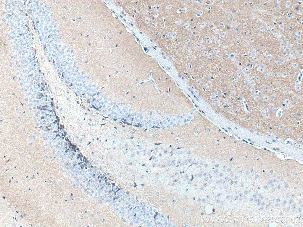
IHC staining of mouse brain using 66219-1-Ig (same clone as 66219-1-PBS)
Immunohistochemical analysis of paraffin-embedded mouse brain tissue slide using 66219-1-Ig (N-cadherin antibody) at dilution of 1:10000 (under 10x lens. Heat mediated antigen retrieval with Tris-EDTA buffer (pH 9.0). This data was developed using the same antibody clone with 66219-1-PBS in a different storage buffer formulation.
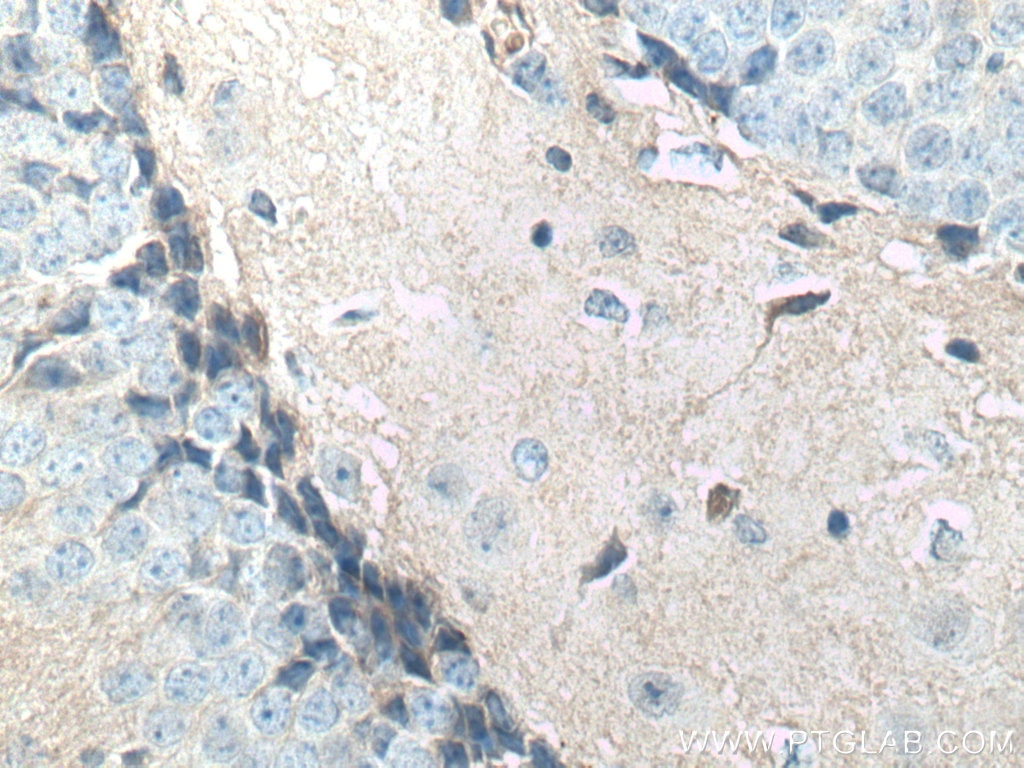
IHC staining of mouse brain using 66219-1-Ig (same clone as 66219-1-PBS)
Immunohistochemical analysis of paraffin-embedded mouse brain tissue slide using 66219-1-Ig (N-cadherin antibody) at dilution of 1:10000 (under 40x lens. Heat mediated antigen retrieval with Tris-EDTA buffer (pH 9.0). This data was developed using the same antibody clone with 66219-1-PBS in a different storage buffer formulation.
IHC-AUTOSTAINER Figures
IHC staining of human heart using 66219-1-Ig (same clone as 66219-1-PBS)
Immunohistochemical analysis of paraffin-embedded human heart tissue slide using 66219-1-Ig (N-cadherin antibody) at dilution of 1:6000. Heat mediated antigen retrieval with Tris-EDTA buffer (pH 9.0). The staining was performed on an autostainer. This data was developed using the same antibody clone with 66219-1-PBS in a different storage buffer formulation.
IHC staining of human brain using 66219-1-Ig (same clone as 66219-1-PBS)
Immunohistochemical analysis of paraffin-embedded human brain tissue slide using 66219-1-Ig (N-cadherin antibody) at dilution of 1:6000 (under 40x lens). Heat mediated antigen retrieval with Tris-EDTA buffer (pH 9.0). The staining was performed on an autostainer. This data was developed using the same antibody clone with 66219-1-PBS in a different storage buffer formulation.
IHC staining of human liver using 66219-1-Ig (same clone as 66219-1-PBS)
Immunohistochemical analysis of paraffin-embedded human liver tissue slide using 66219-1-Ig (N-cadherin antibody) at dilution of 1:6000. Heat mediated antigen retrieval with Tris-EDTA buffer (pH 9.0). The staining was performed on an autostainer. This data was developed using the same antibody clone with 66219-1-PBS in a different storage buffer formulation.
IF-P Figures
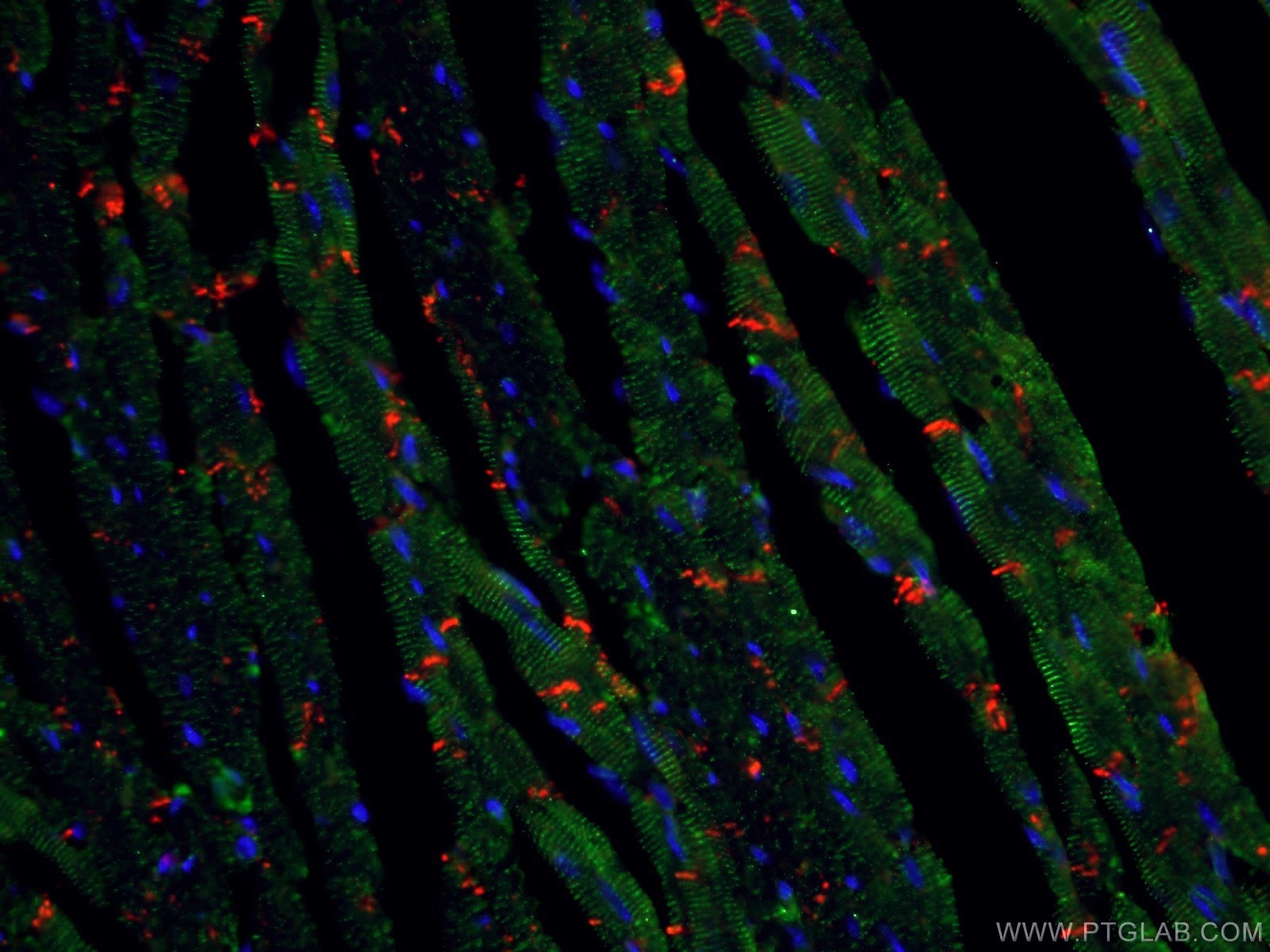
IF Staining of mouse heart using 66219-1-Ig (same clone as 66219-1-PBS)
Immunofluorescent analysis of (4% PFA) fixed mouse heart tissue using 66219-1-Ig (N-cadherin antibody) at dilution of 1:100 and CoraLite594-Conjugated AffiniPure Goat Anti-Mouse IgG(H+L). The section was co-stained with 11313-2-AP (alpha Actinin) in green. This data was developed using the same antibody clone with 66219-1-PBS in a different storage buffer formulation.
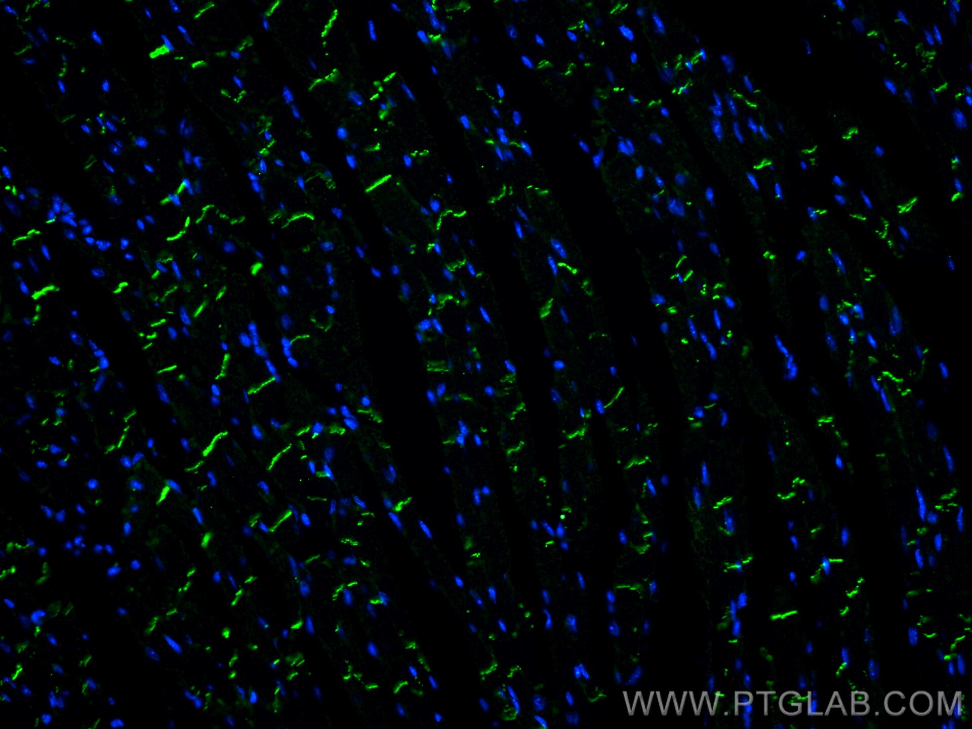
IF Staining of mouse heart using 66219-1-Ig (same clone as 66219-1-PBS)
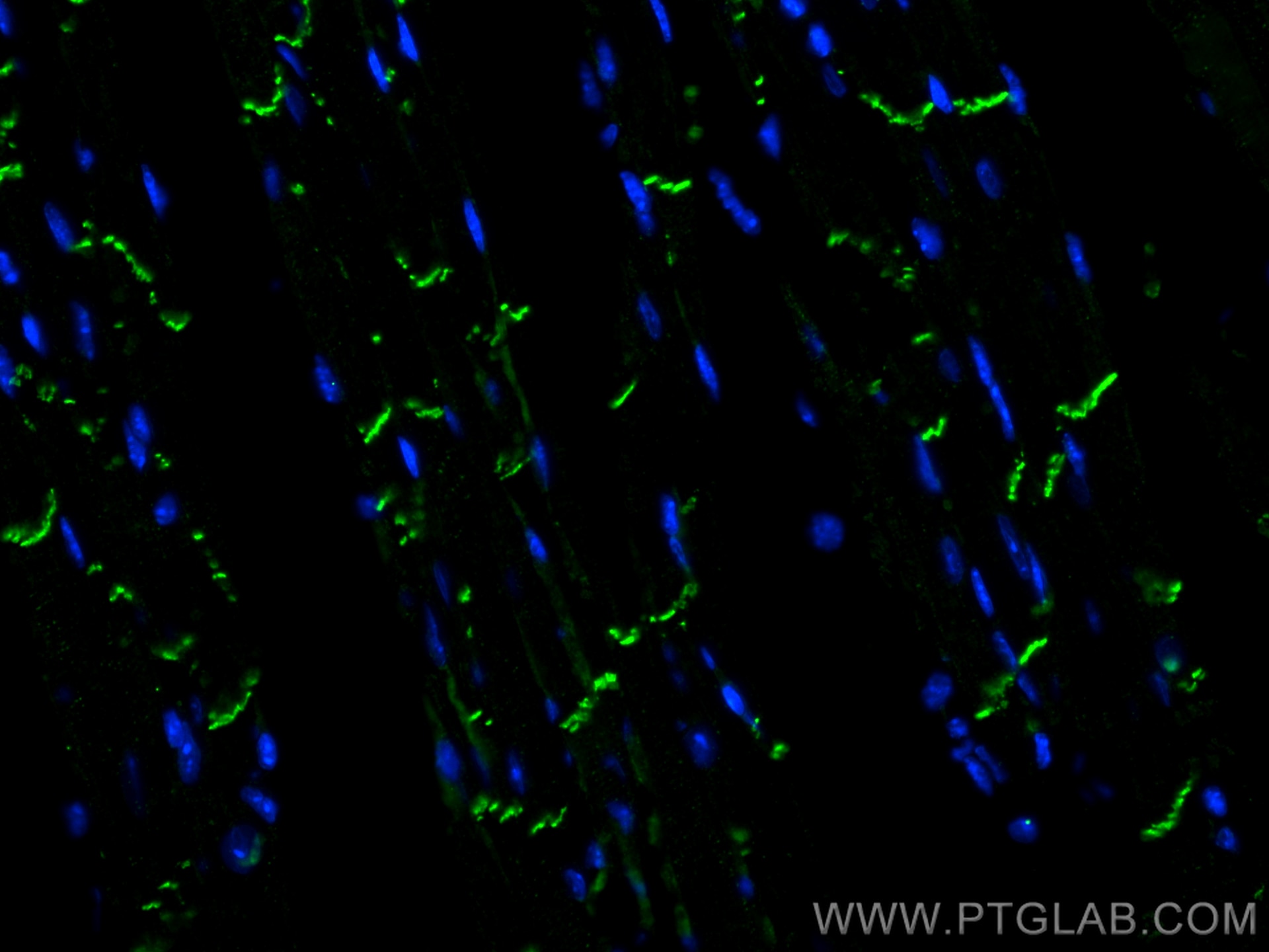
Immunofluorescent analysis of (4% PFA) fixed paraffin-embedded mouse heart tissue using N-cadherin antibody (66219-1-Ig, Clone: 1D8B3 ) at dilution of 1:400 and CoraLite®488-Conjugated Goat Anti-Mouse IgG(H+L) (SA00013-1). Heat mediated antigen retrieval with Tris-EDTA buffer (pH 9.0). This data was developed using the same antibody clone with 66219-1-PBS in a different storage buffer formulation.
CYTOMETRIC BEAD ARRAY Figures
Cytometric bead array standard curve of MP50439-2
Cytometric bead array standard curve of MP50439-2, N-cadherin Monoclonal Matched Antibody Pair, PBS Only. Capture antibody: 66219-1-PBS. Detection antibody: 66219-2-PBS. Standard:Eg0701. Range: 0.781-100 ng/mL.