
Recombinant Mouse CD28 protein (hFc Tag)
Species
Mouse
Purity
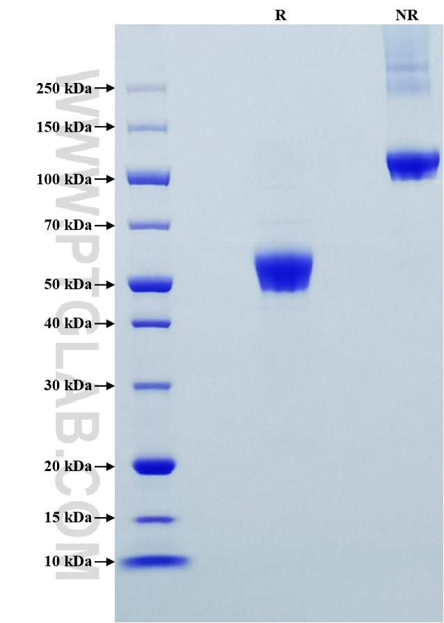
>95 %, SDS-PAGE
Tag
hFc Tag
Activity
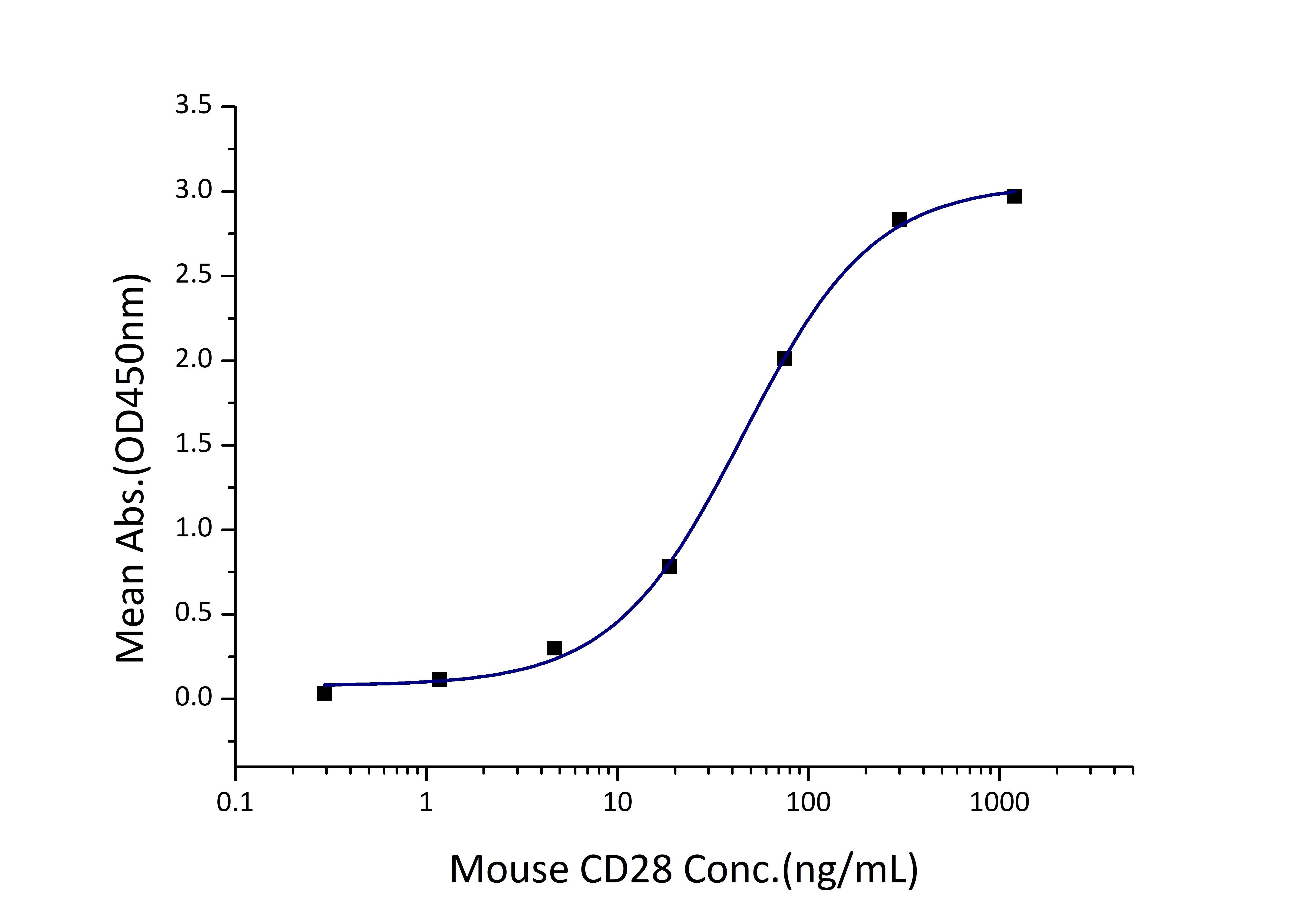
EC50: 23-91 ng/mL
Cat no : Eg0805
Validation Data Gallery
Product Information
| Purity | >95 %, SDS-PAGE |
| Endotoxin | <0.1 EU/μg protein, LAL method |
| Activity |
Immobilized Mouse CD86 (His tag) at 1 μg/mL (100 μL/well) can bind Mouse CD28 (hFc tag) with a linear range of 23-91 ng/mL. |
| Expression | HEK293-derived Mouse CD28 protein Asn20-Leu150 (Accession# P31041) with a Human IgG1 Fc tag at the C-terminus. |
| GeneID | 12487 |
| Accession | P31041 |
| PredictedSize | 41.5 kDa |
| SDS-PAGE | 48-60 kDa, reducing (R) conditions |
| Formulation | Lyophilized from 0.22 μm filtered solution in PBS, pH 7.4. Normally 5% trehalose and 5% mannitol are added as protectants before lyophilization. |
| Reconstitution | Briefly centrifuge the tube before opening. Reconstitute at 0.1-0.5 mg/mL in sterile water. |
| Storage Conditions |
It is recommended that the protein be aliquoted for optimal storage. Avoid repeated freeze-thaw cycles.
|
| Shipping | The product is shipped at ambient temperature. Upon receipt, store it immediately at the recommended temperature. |
Background
CD28 (T-cell-specific surface glycoprotein CD28), also known as T44 and Tp44, is a 44 kD disulfide-linked homodimeric type I glycoprotein. It is a member of the immunoglobulin superfamily and is expressed on most T lineage cells, NK cell subsets, and plasma cells. CD28 may affect in vivo immune responses by functioning both as a cell adhesion molecule linking B and T lymphocytes and as the surface component of a novel signal transduction pathway. CD28 binds both CD80 and CD86 with a highly conserved motif MYPPY in the CDR3-like loop. CD28 is considered a major co-stimulatory molecule, inducing T lymphocyte activation and IL-2 synthesis, and preventing cell death.
References:
1.W Lesslauer, et al. (1986) Eur J Immunol. 16(10):1289-96.
2.C H June, et al. (1990) Immunol Today. 11(6):211-6.
3.T M Williams, et al. (1992) J Immunol. 148(8):2609-16.
4.P S Linsley, et al. (1993) Annu Rev Immunol. 11:191-212.
5.R J Peach, et al. (1994) J Exp Med. 180(6):2049-58.
6.Edward J Evans, et al. (2005) Nat Immunol. 6(3):271-9.