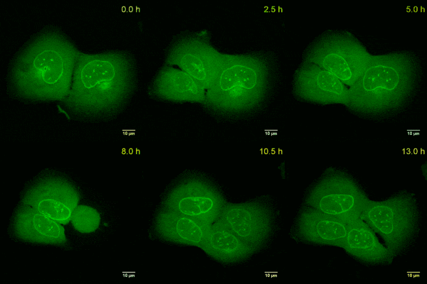
Validation Data Gallery
Product Information
DNA plasmid encoding for anti-Lamin VHH (anti-Lamin Nanobody) fused to TagGFP2.
| Description | The Lamin-Chromobody visualizes the nuclear lamina. • Easily transfect your cells with the Lamin-Chromobody plasmid • Visualize the nuclear lamina without interfering with its cellular functions • Monitor the nuclear integrity and morphology during live cell microscopy
|
| Applications | Trace the nuclear lamina in live cells in real-time Monitor the nuclear integrity and morphology Use the Lamin-Chromobody® as a biosensor for real-time assays of mitosis or apoptosis |
| Specificity/Target | Lamin A/C, tested in human cell lines (HeLa, U2OS) and insect S2 cells. |
| Reporter | TagGFP2 |
| Vector type | Mammalian expression vector |
| Promoter | Constitutive CMV IE |
| Codon usage | Mammalian |
| Selection | Kan/Neo |
| Sequence | With the Lamin-Chromobody plasmid you receive the sequence information of the Alpaca antibody to Lamin fused to TagGFP2, as well as the full vector sequence. |
| Transfection | Transfection of Chromobody plasmids into mammalian cells can be done with standard DNA-transfection methods, e.g. lipofection (Lipofectamine 2000® from Thermo Scientific), according to the manufacturer’s protocol for the transfection reagent. Please choose the transfection method that works the best for your cell type. |
| Microscopy techniques | Wide-field epifluorescence microscopy; confocal microscopy |
| Storage Condition | Shipped at ambient temperature. Store at -20 °C |
Documentation
| SDS |
|---|
| lcg_SDS_Lamin-Chromobody® plasmid (TagGFP2) (EN) |
| Datasheet |
|---|
| Lamin-Chromobody® plasmid (TagGFP2) Datasheet |
| EULA |
|---|
| lcg End User License Agreement (EULA) Lamin Chromobody TagGFP plasmid non-profit (US) (PDF) |
| lcg End User License Agreement (EULA) Lamin Chromobody TagGFP plasmid non-profit (PDF) |
| Brochure |
|---|
| Chromotek nanobodies brochure (PDF) |
Publications
| Application | Title |
|---|---|
Nat Cell Biol A transient pool of nuclear F-actin at mitotic exit controls chromatin organization. | |
Nat Commun GPCR-induced calcium transients trigger nuclear actin assembly for chromatin dynamics. | |
PLoS Pathog Fluorescent protein tagging of adenoviral proteins pV and pIX reveals 'late virion accumulation compartment'. | |
Sci Rep A novel epitope tagging system to visualize and monitor antigens in live cells with chromobodies. | |
Cell Mol Bioeng Characterization of 3D Printed Stretching Devices for Imaging Force Transmission in Live-Cells. |


