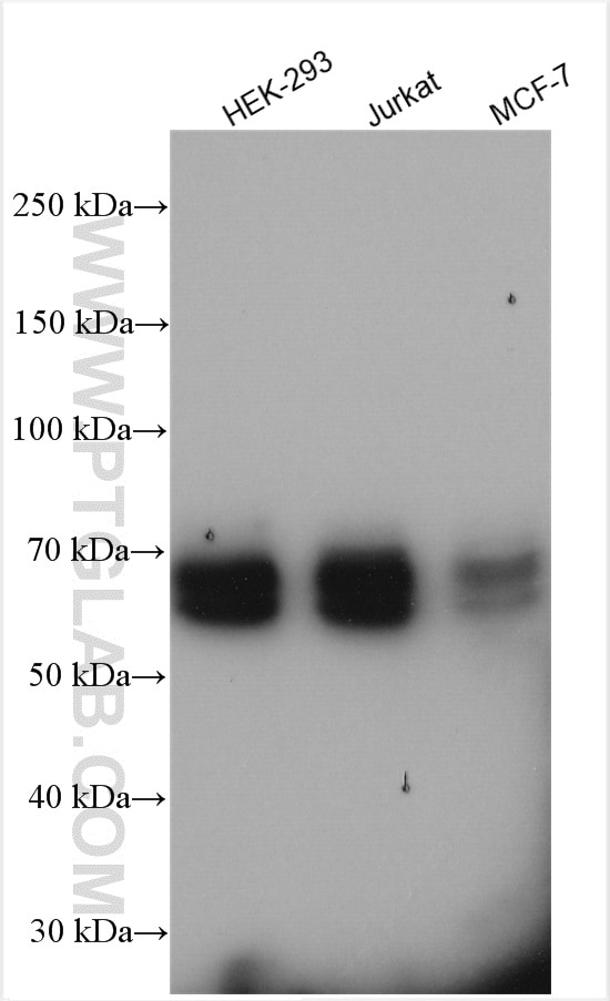
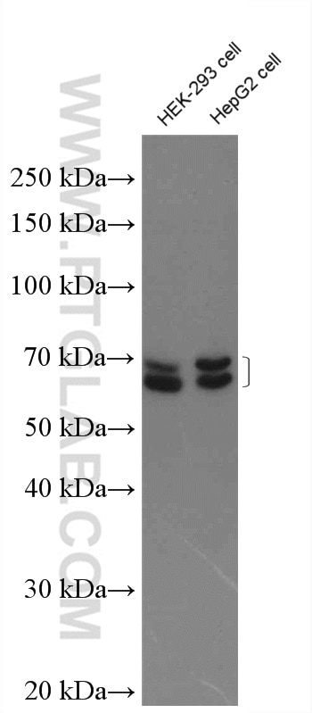
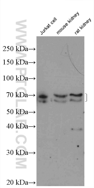
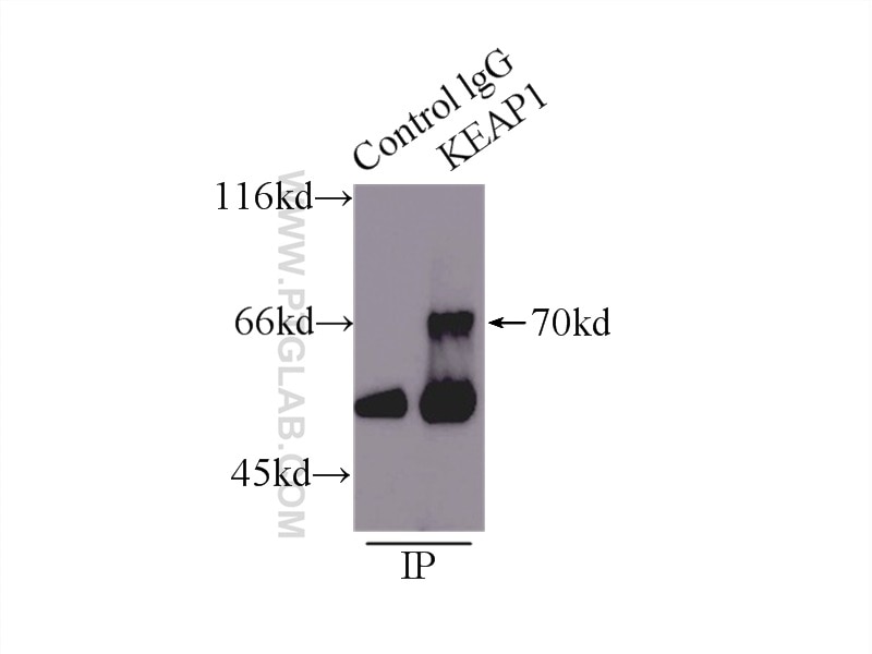
Validation Data Gallery
Tested Applications
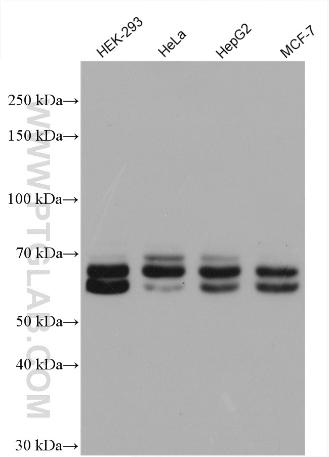
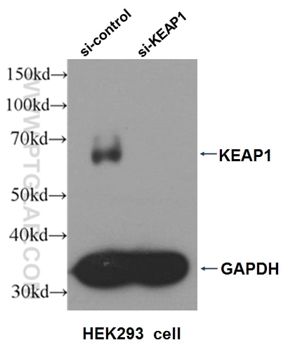
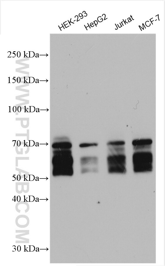
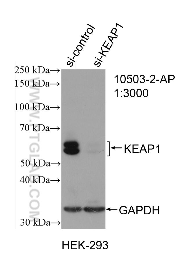
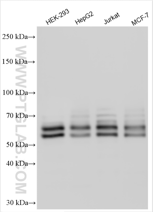
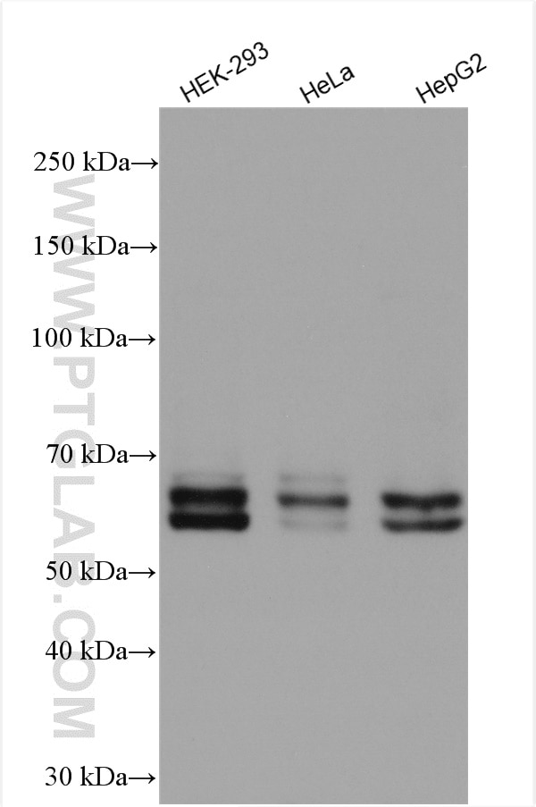
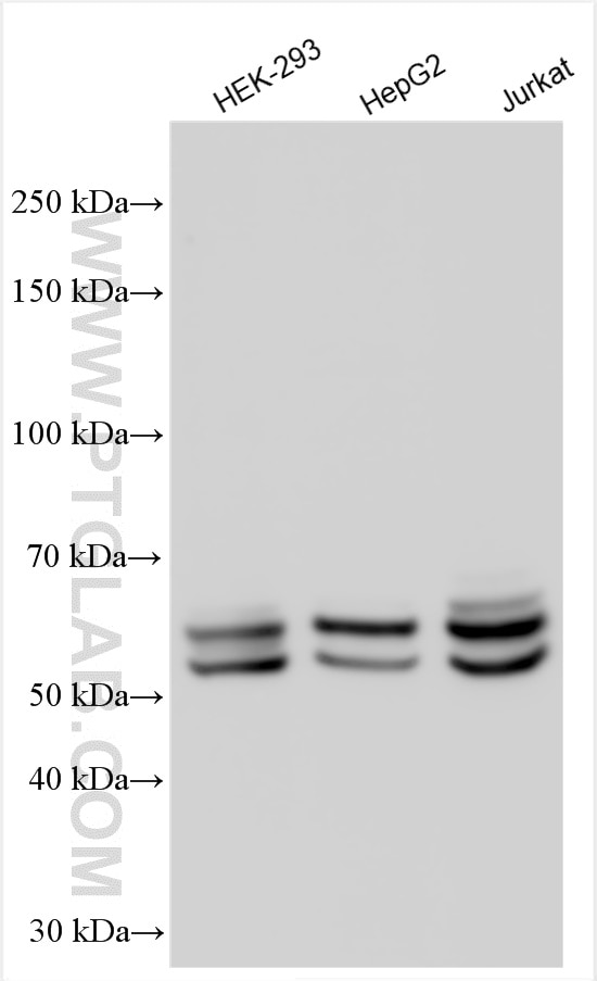
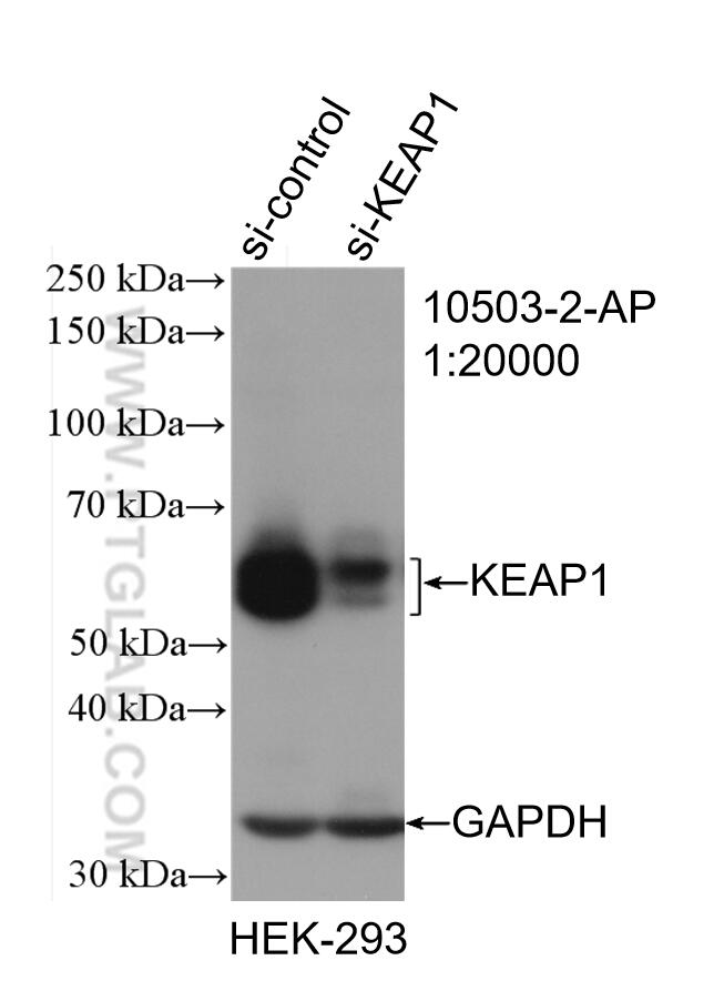
| Positive WB detected in | HEK-293 cells, HepG2 cells, Jurkat cells, MCF-7 cells, HeLa cells |
| Positive IP detected in | mouse skeletal muscle tissue |
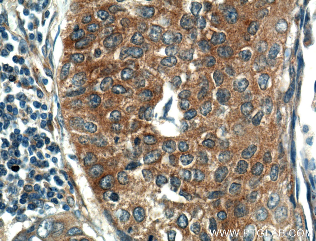
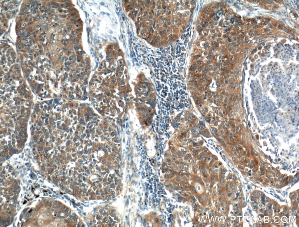
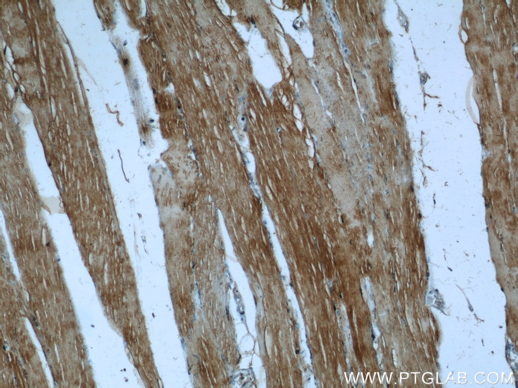
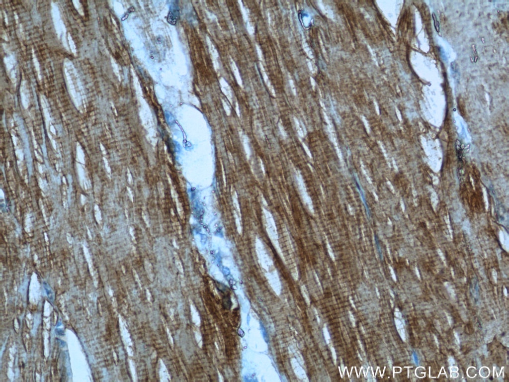
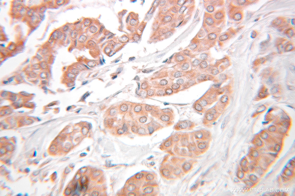
| Positive IHC detected in | human lung cancer tissue, human breast cancer tissue, human skeletal muscle tissue Note: suggested antigen retrieval with TE buffer pH 9.0; (*) Alternatively, antigen retrieval may be performed with citrate buffer pH 6.0 |
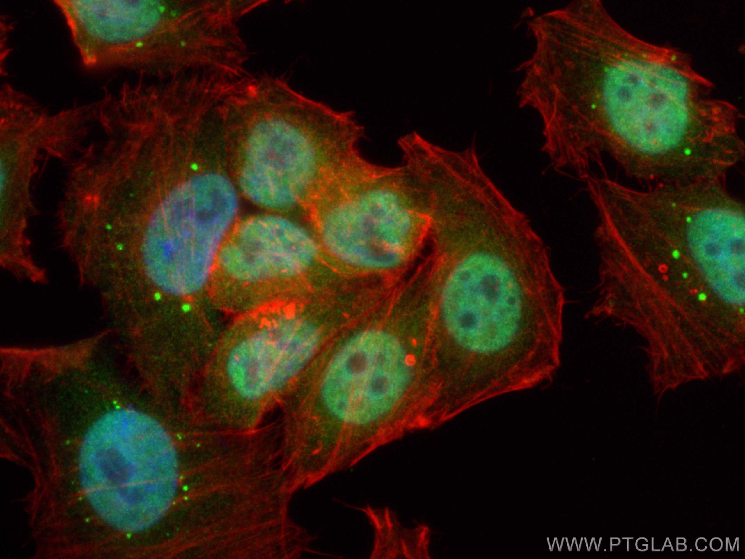
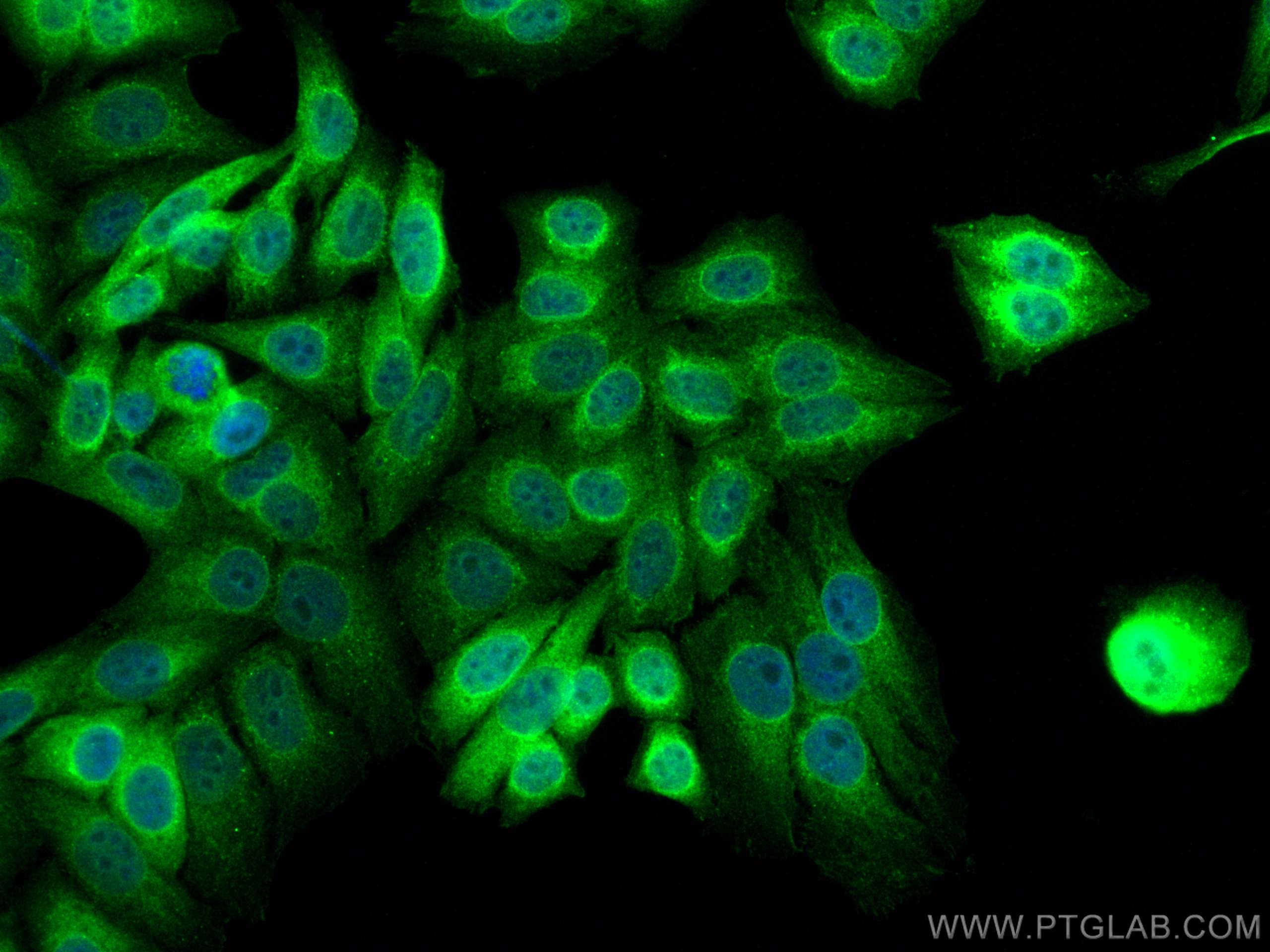
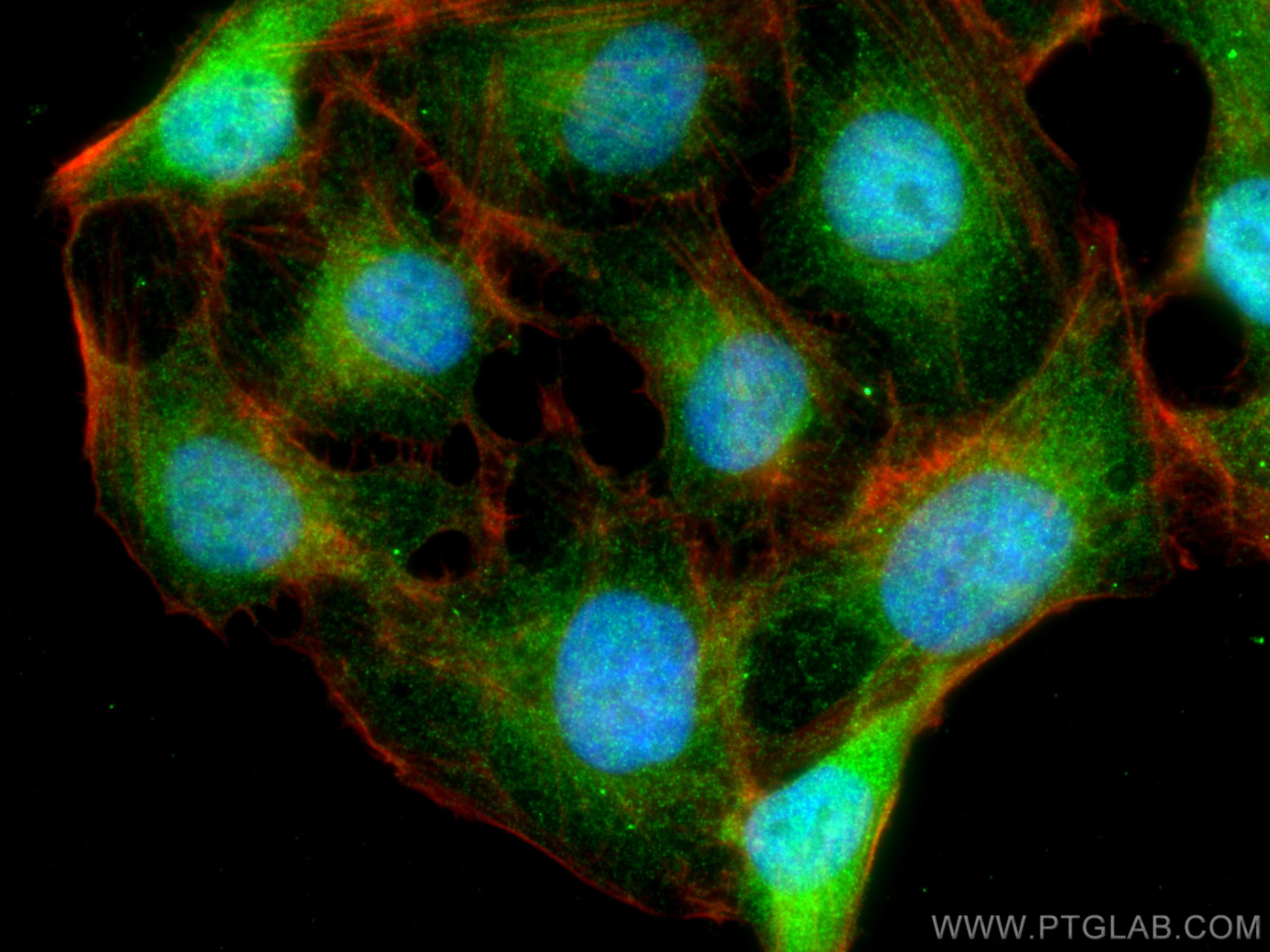
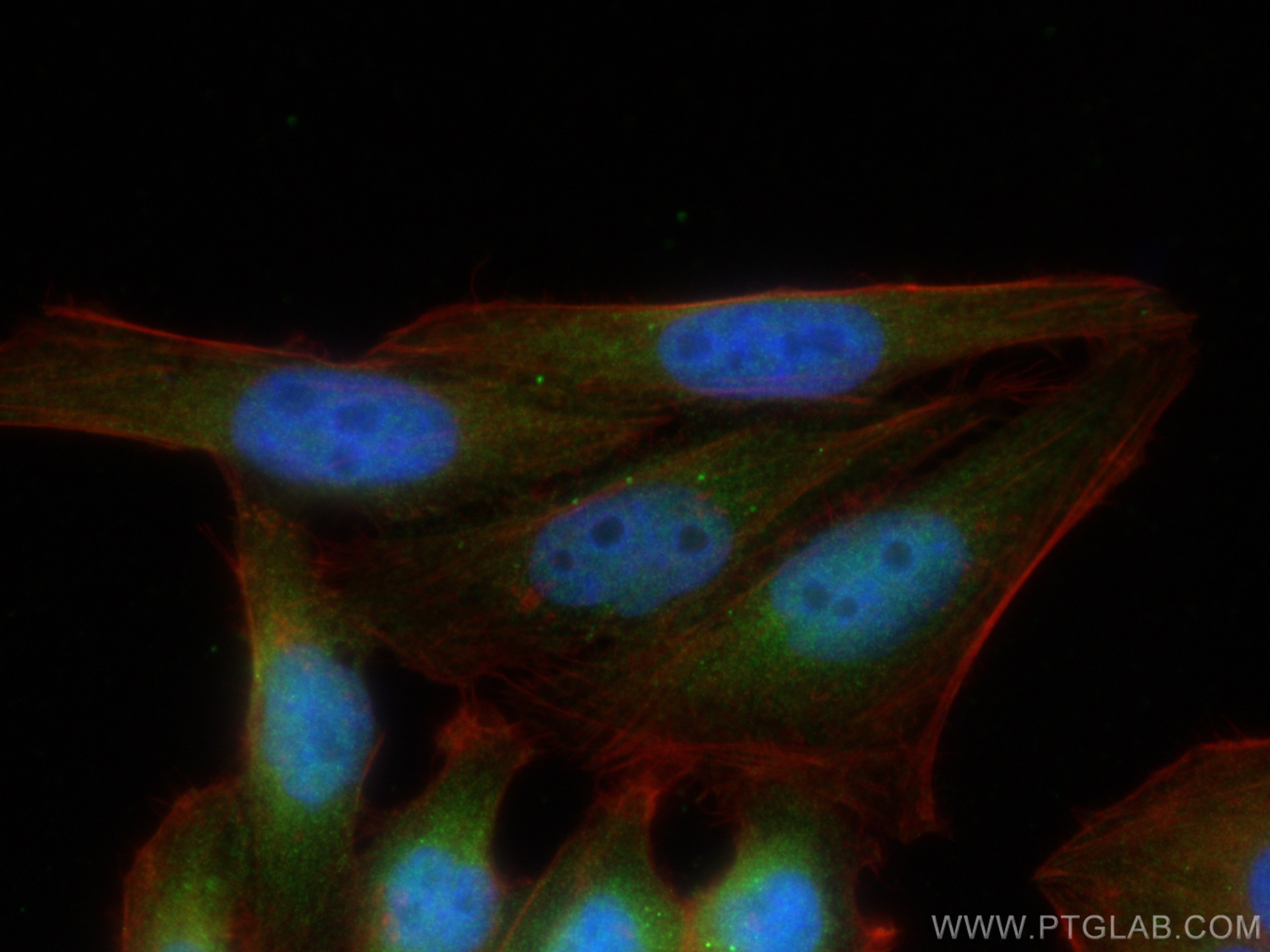
| Positive IF/ICC detected in | U2OS cells, HepG2 cells |
Recommended dilution
| Application | Dilution |
|---|---|
| Western Blot (WB) | WB : 1:2000-1:10000 |
| Immunoprecipitation (IP) | IP : 0.5-4.0 ug for 1.0-3.0 mg of total protein lysate |
| Immunohistochemistry (IHC) | IHC : 1:50-1:500 |
| Immunofluorescence (IF)/ICC | IF/ICC : 1:50-1:500 |
| It is recommended that this reagent should be titrated in each testing system to obtain optimal results. | |
| Sample-dependent, Check data in validation data gallery. | |
Product Information
10503-2-AP targets KEAP1 in WB, IHC, IF/ICC, IP, CoIP, RIP, ELISA applications and shows reactivity with human, mouse samples.
| Tested Reactivity | human, mouse |
| Cited Reactivity | human, mouse, rat, pig, monkey, chicken, bovine, yellow catfish |
| Host / Isotype | Rabbit / IgG |
| Class | Polyclonal |
| Type | Antibody |
| Immunogen |
CatNo: Ag0779 Product name: Recombinant human KEAP1 protein Source: e coli.-derived, PGEX-4T Tag: GST Domain: 325-624 aa of BC002930 Sequence: GRLIYTAGGYFRQSLSYLEAYNPSDGTWLRLADLQVPRSGLAGCVVGGLLYAVGGRNNSPDGNTDSSALDCYNPMTNQWSPCAPMSVPRNRIGVGVIDGHIYAVGGSHGCIHHNSVERYEPERDEWHLVAPMLTRRIGVGVAVLNRLLYAVGGFDGTNRLNSAECYYPERNEWRMITAMNTIRSGAGVCVLHNCIYAAGGYDGQDQLNSVERYDVETETWTFVAPMKHRRSALGITVHQGRIYVLGGYDGHTFLDSVECYDPDTDTWSEVTRMTSGRSGVGVAVTMEPCRKQIDQQNCTC 相同性解析による交差性が予測される生物種 |
| Full Name | kelch-like ECH-associated protein 1 |
| Calculated molecular weight | 624 aa, 70 kDa |
| Observed molecular weight | 55~70 kDa |
| GenBank accession number | BC002930 |
| Gene Symbol | KEAP1 |
| Gene ID (NCBI) | 9817 |
| RRID | AB_2132625 |
| Conjugate | Unconjugated |
| Form | |
| Form | Liquid |
| Purification Method | Antigen affinity purification |
| UNIPROT ID | Q14145 |
| Storage Buffer | PBS with 0.02% sodium azide and 50% glycerol{{ptg:BufferTemp}}7.3 |
| Storage Conditions | Store at -20°C. Stable for one year after shipment. Aliquoting is unnecessary for -20oC storage. |
Background Information
Kelch-like ECH-associated protein 1 (KEAP1) is a negative regulator of nuclear factor erythroid 2-related factor 2 (Nrf2), a transcription factor governing the antioxidant response.
What is the molecular weight of KEAP1 protein? Are there any isoforms of KEAP1?
The molecular weight of KEAP1 protein is 70 kDa. The KEAP1 gene gives rise only to protein isoforms, but mutations of KEAP1 protein have been found in various cancer types.
What is the subcellular localization of KEAP1?
KEAP1 resides in the cytoplasm, where it binds to Nrf2, targeting it for degradation and preventing translocation of Nrf2 to the nucleus.
How does KEAP1 control Nrf2 levels? Is KEAP1 post-translationally modified?
KEAP1 is rich in reactive cysteine residues, whose thiol groups play a role in binding to CUL3 and the polyubiquitination of Nrf2, which leads to degradation of Nrf2 via the proteasome system. During oxidative stress, electrophiles and reactive oxygen species (ROS) modify the KEAP1 thiol groups, reducing the affinity of KEAP1 to CUL3 and the stabilization of Nrf2. Nrf2 then translocates to the nucleus, where it binds to the antioxidant responsive elements (AREs) and induces the expression of antioxidant proteins (PMID: 16354693).
How to measure oxidative stress using KEAP1 and Nrf2 proteins as a readout
Under basal conditions (unstressed cells), a detectable KEAP1 protein level is observed. Oxidative stress modifies KEAP1 protein activity by increasing the Nrf2 protein levels. This can be measured, for example, using western blotting (PMID: 27697860). KEAP1 protein levels are not altered by oxidative stress.
What is the role of the KEAP1-Nrf2 pathway in health and disease?
The KEAP-Nrf2 pathway plays a vital role in redox homeostasis and cryoprotection. Inhibition of KEAP1 activity leads to the activation of Nrf2 and increase the response to oxidative stress and anti-inflammatory effects (PMID: 29717933). The activation of Nrf2 can be beneficial in the case of metabolic diseases, such as diabetes, as well as neurodegenerative diseases such as Parkinson's and Alzheimer's diseases. However, the increased activation of Nrf2 is also known to promote tumor growth and metastasis. Mutations in both KEAP1 and Nrf2 were found in various solid tumor types.
Protocols
| Product Specific Protocols | |
|---|---|
| IF protocol for KEAP1 antibody 10503-2-AP | Download protocol |
| IHC protocol for KEAP1 antibody 10503-2-AP | Download protocol |
| IP protocol for KEAP1 antibody 10503-2-AP | Download protocol |
| WB protocol for KEAP1 antibody 10503-2-AP | Download protocol |
| Standard Protocols | |
|---|---|
| Click here to view our Standard Protocols |
Publications
| Species | Application | Title |
|---|---|---|
Cell Recognition of BACH1 quaternary structure degrons by two F-box proteins under oxidative stress | ||
Nature KLHL22 activates amino-acid-dependent mTORC1 signalling to promote tumorigenesis and ageing.
| ||
Cell Systematic discovery of mutation-directed neo-protein-protein interactions in cancer.
| ||
Cell Nrf2 Activation Promotes Lung Cancer Metastasis by Inhibiting the Degradation of Bach1. | ||
Mol Cancer Double vulnerability of active-NRF2 lung squamous cell carcinoma to NRF2 and TRIM24 |

