Recombinant Human FUT4 protein (rFc Tag)
Species
Human
Purity
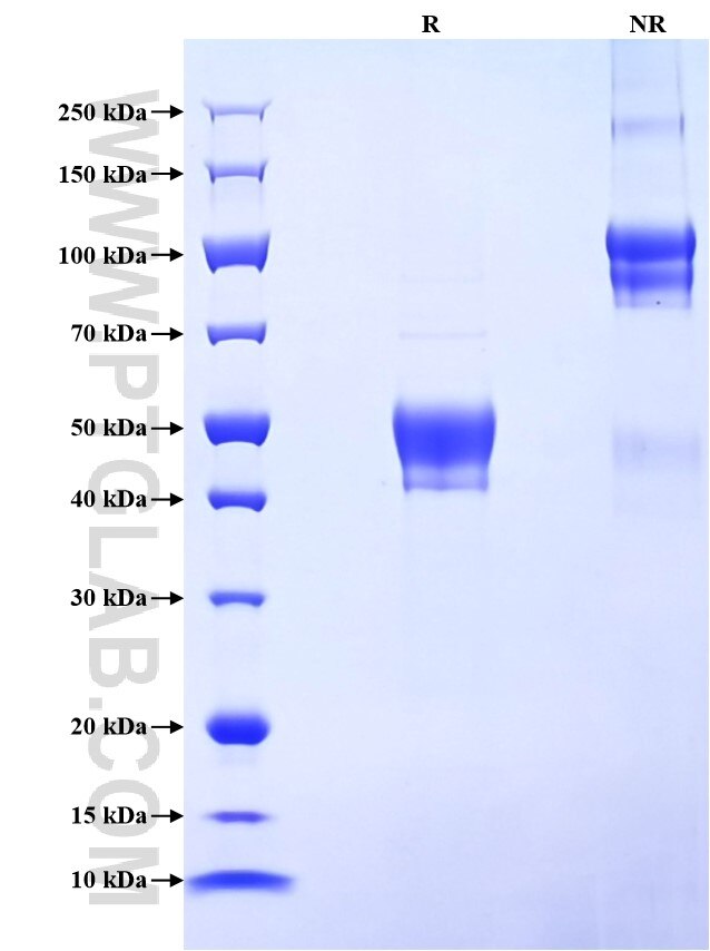
>90 %, SDS-PAGE
Tag
rFc Tag
Activity
not tested
Cat no : Eg2194
Validation Data Gallery
Product Information
| Purity | >90 %, SDS-PAGE |
| Endotoxin | <0.1 EU/μg protein, LAL method |
| Activity |
Not tested |
| Expression | HEK293-derived Human FUT4 protein Gly199-His302 (Accession# P22083-1) with a rabbit IgG Fc tag at the C-terminus. |
| GeneID | 2526 |
| Accession | P22083-1 |
| PredictedSize | 37.8 kDa |
| SDS-PAGE | 42-55 kDa, reducing (R) conditions |
| Formulation | Lyophilized from 0.22 μm filtered solution in PBS, pH 7.4. Normally 5% trehalose and 5% mannitol are added as protectants before lyophilization. |
| Reconstitution | Briefly centrifuge the tube before opening. Reconstitute at 0.1-0.5 mg/mL in sterile water. |
| Storage Conditions |
It is recommended that the protein be aliquoted for optimal storage. Avoid repeated freeze-thaw cycles.
|
| Shipping | The product is shipped at ambient temperature. Upon receipt, store it immediately at the recommended temperature. |
Background
FUT4, also named as ELFT and FCT3A, belongs to the glycosyltransferase 10 family. FUT4 may catalyze alpha-1,3 glycosidic linkages involved in the expression of Lewis X/SSEA-1 and VIM-2 antigens. The expression of CD15 (acts as a terminal glycotope in glycoproteins and glycolipids) is directed by FUT4 in promyelocytes and monocytes. FUT4 is an antigenic epitope defined as a Lewis X carbohydrate structure is expressed on murine embryonal carcinoma cells (EC), murine ES and iPS cells, and murine and human germ cells. It is widely used as a positive surface marker for mouse undifferentiated ES and iPS cells and a negative surface marker for human undifferentiated ES and iPS cells. Expression is down-regulated following differentiation of murine EC and ES cells, while the differentiation of human EC and ES cells is accompanied by an increase in FUT4 expression. FUT4 is associated with cell adhesion, migration and differentiation. 19497-1-AP antibody detects the glycosylated isoform proteins around 95-140 kDa in SDS-PAGE.
References:
1. Yu, Ming et al. Scientific reports vol. 7,1 5315.
2. Zheng, Qin et al. Cell death and differentiation vol. 24,12 (2017): 2161-2172.
3. Nakayama, F et al. The Journal of biological chemistry vol. 276,19 (2001): 16100-6.
