WB Figures
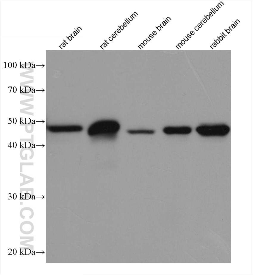
WB analysis using 60190-1-Ig (same clone as 60190-1-PBS)
Various lysates were subjected to SDS PAGE followed by western blot with 60190-1-Ig (GFAP antibody) at dilution of 1:100000 incubated at room temperature for 1.5 hours. This data was developed using the same antibody clone with 60190-1-PBS in a different storage buffer formulation.
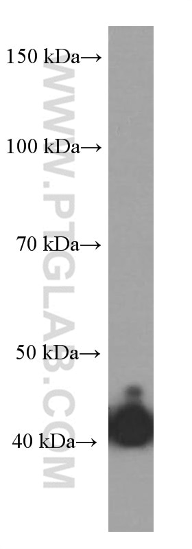
WB analysis of U-251 using 60190-1-Ig (same clone as 60190-1-PBS)
U-251 cells were subjected to SDS PAGE followed by western blot with 60190-1-Ig (GFAP Antibody) at dilution of 1:1500 incubated at room temperature for 1.5 hours. This data was developed using the same antibody clone with 60190-1-PBS in a different storage buffer formulation.
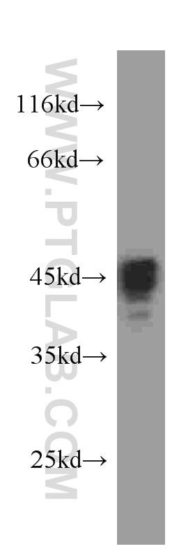
WB analysis of human brain using 60190-1-Ig (same clone as 60190-1-PBS)
human brain tissue were subjected to SDS PAGE followed by western blot with 60190-1-Ig (GFAP antibody) at dilution of 1:10000 incubated at room temperature for 1.5 hours. This data was developed using the same antibody clone with 60190-1-PBS in a different storage buffer formulation.
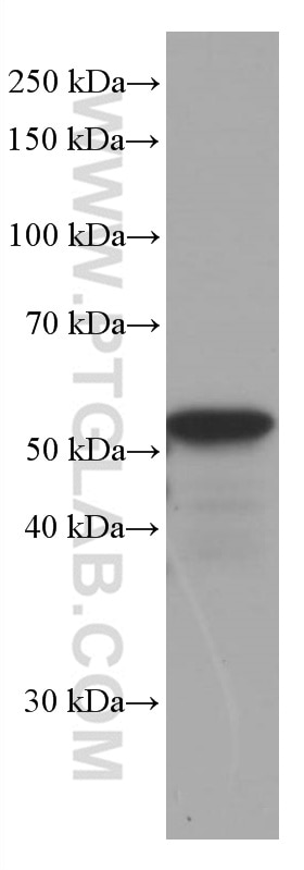
WB analysis of pig brain using 60190-1-Ig (same clone as 60190-1-PBS)
pig brain tissue were subjected to SDS PAGE followed by western blot with 60190-1-Ig (GFAP antibody) at dilution of 1:300000 incubated at room temperature for 1.5 hours. This data was developed using the same antibody clone with 60190-1-PBS in a different storage buffer formulation.
IHC Figures
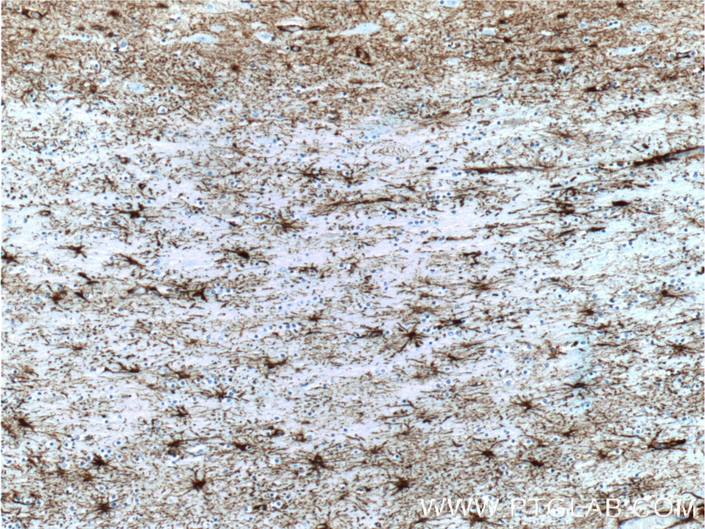
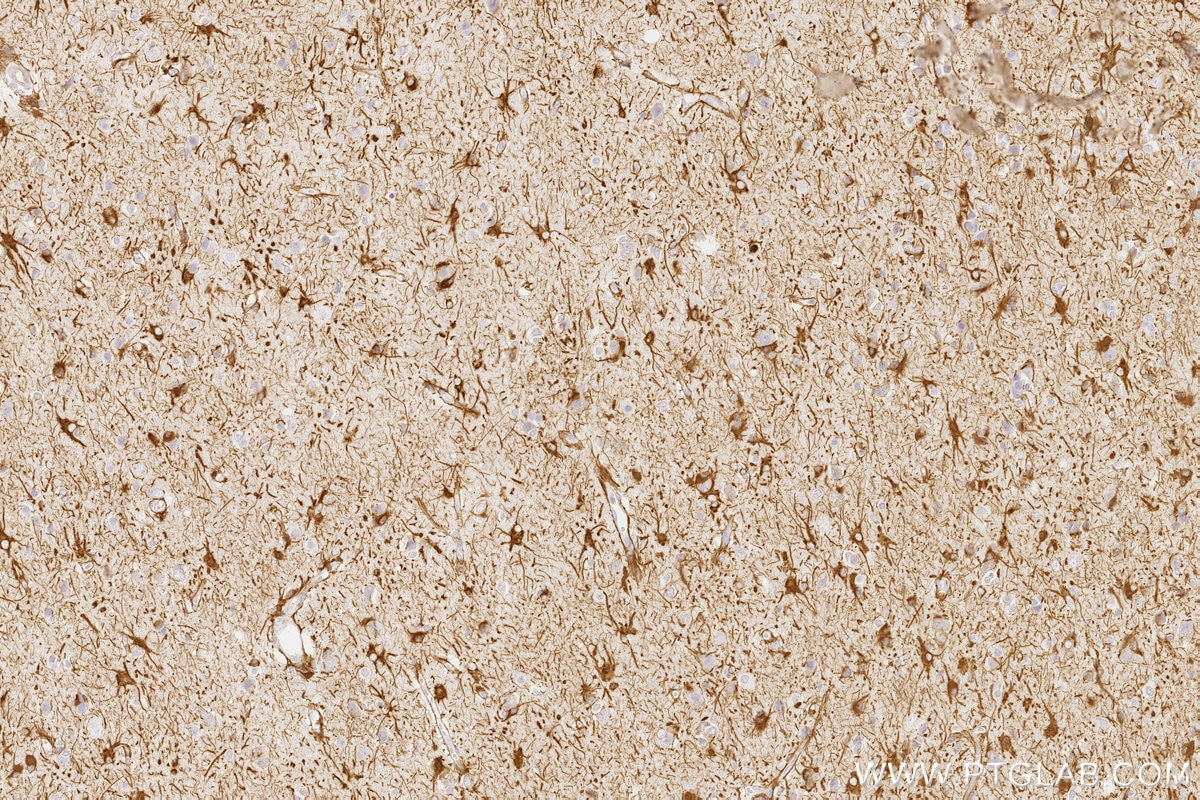
IHC staining of human brain using 60190-1-Ig (same clone as 60190-1-PBS)
Immunohistochemical analysis of paraffin-embedded human brain tissue slide using 60190-1-Ig (GFAP Antibody) at dilution of 1:5000 (under 10x lens). Heat mediated antigen retrieved with Citric acid buffer, pH6.0. This data was developed using the same antibody clone with 60190-1-PBS in a different storage buffer formulation.
IHC staining of mouse brain using 60190-1-Ig (same clone as 60190-1-PBS)
Immunohistochemical analysis of paraffin-embedded mouse brain tissue slide using 60190-1-Ig (GFAP antibody) at dilution of 1:5000 (under 10x lens). Heat mediated antigen retrieval with Tris-EDTA buffer (pH 9.0). This data was developed using the same antibody clone with 60190-1-PBS in a different storage buffer formulation.
IHC staining of mouse brain using 60190-1-Ig (same clone as 60190-1-PBS)
Immunohistochemical analysis of paraffin-embedded mouse brain tissue slide using 60190-1-Ig (GFAP antibody) at dilution of 1:5000 (under 10x lens). Heat mediated antigen retrieval with Tris-EDTA buffer (pH 9.0). This data was developed using the same antibody clone with 60190-1-PBS in a different storage buffer formulation.
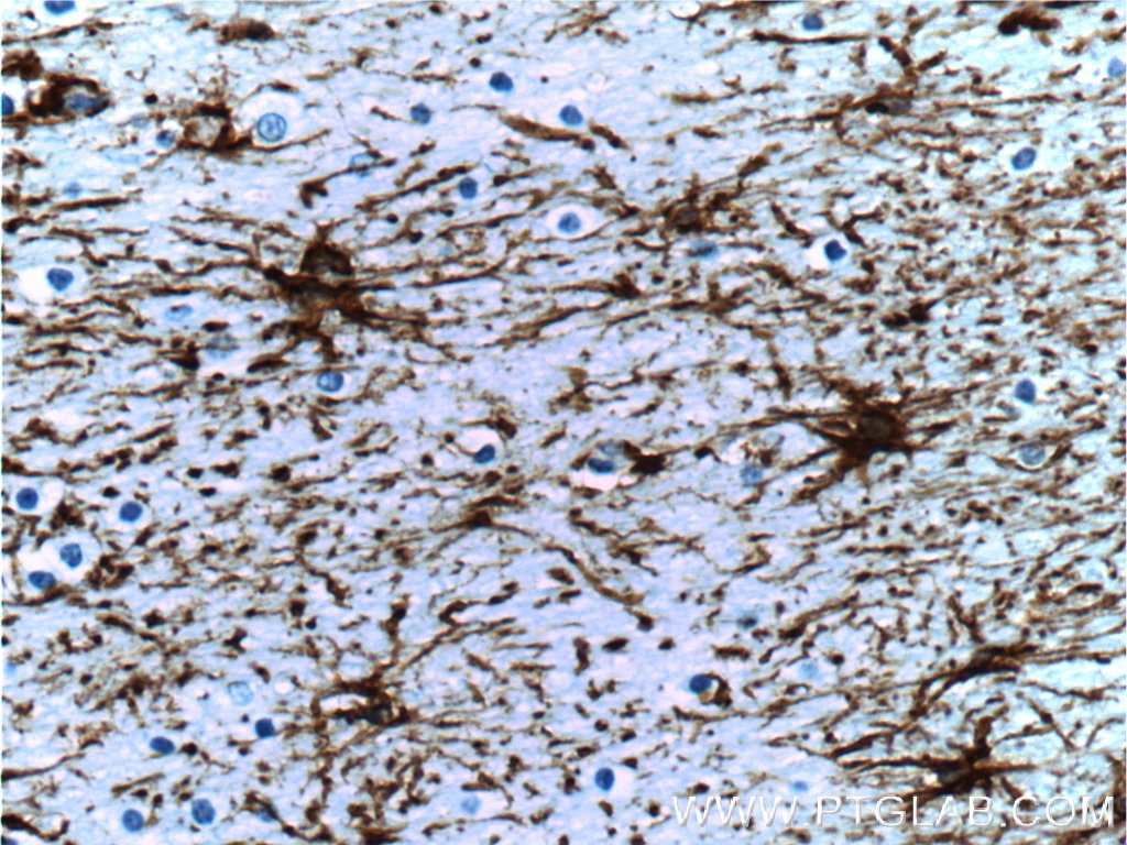
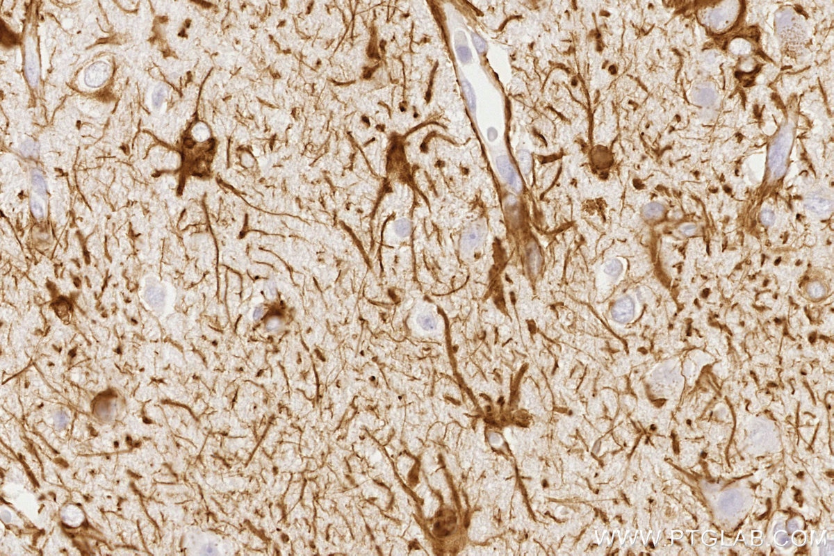
IHC staining of human brain using 60190-1-Ig (same clone as 60190-1-PBS)
Immunohistochemical analysis of paraffin-embedded human brain tissue slide using 60190-1-Ig (GFAP Antibody) at dilution of 1:5000 (under 40x lens). Heat mediated antigen retrieved with Citric acid buffer, pH6.0. This data was developed using the same antibody clone with 60190-1-PBS in a different storage buffer formulation.
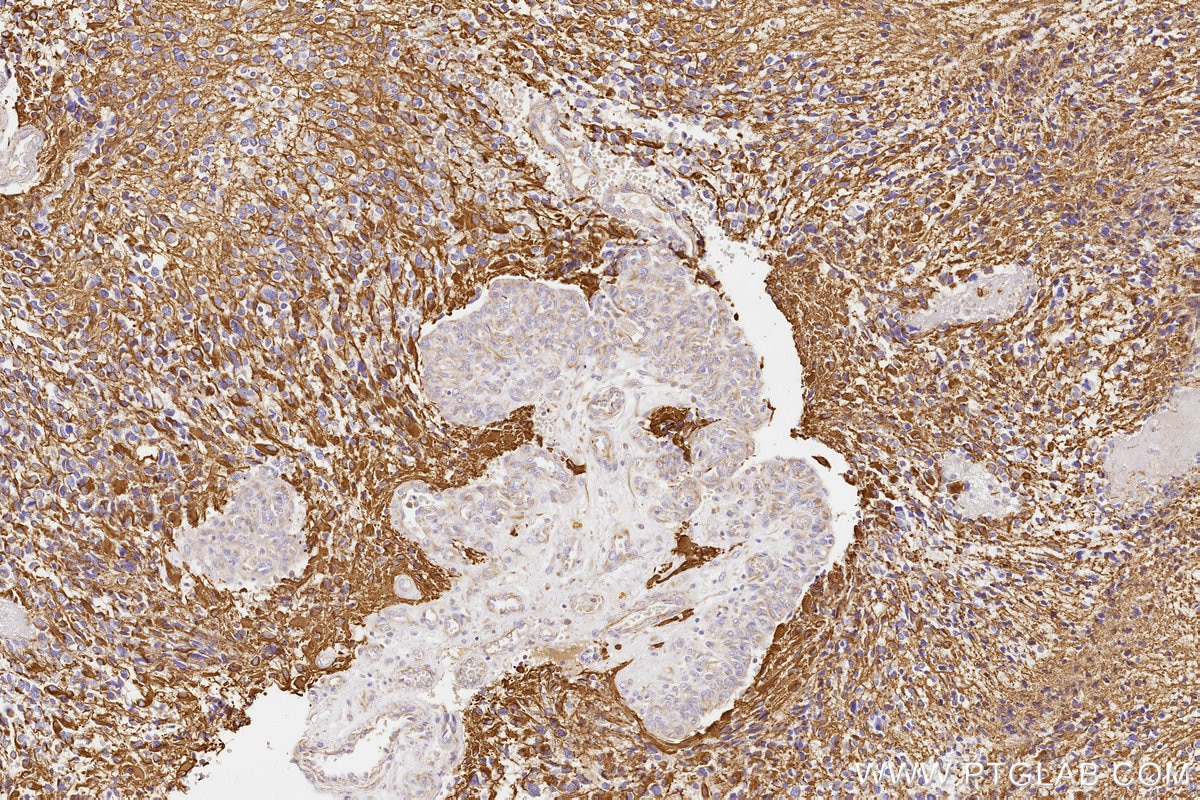
IHC staining of human gliomas using 60190-1-Ig (same clone as 60190-1-PBS)
Immunohistochemical analysis of paraffin-embedded human gliomas tissue slide using 60190-1-Ig (GFAP antibody) at dilution of 1:20000 (under 10x lens). Heat mediated antigen retrieval with Tris-EDTA buffer (pH 9.0). This data was developed using the same antibody clone with 60190-1-PBS in a different storage buffer formulation.
IHC staining of human gliomas using 60190-1-Ig (same clone as 60190-1-PBS)
Immunohistochemical analysis of paraffin-embedded human gliomas tissue slide using 60190-1-Ig (GFAP antibody) at dilution of 1:20000 (under 40x lens). Heat mediated antigen retrieval with Tris-EDTA buffer (pH 9.0). This data was developed using the same antibody clone with 60190-1-PBS in a different storage buffer formulation.
IHC staining of human gliomas using 60190-1-Ig (same clone as 60190-1-PBS)
Immunohistochemical analysis of paraffin-embedded human gliomas tissue slide using 60190-1-Ig (GFAP antibody) at dilution of 1:20000 (under 10x lens). Heat mediated antigen retrieval with Tris-EDTA buffer (pH 9.0). This data was developed using the same antibody clone with 60190-1-PBS in a different storage buffer formulation.
IHC staining of human gliomas using 60190-1-Ig (same clone as 60190-1-PBS)
Immunohistochemical analysis of paraffin-embedded human gliomas tissue slide using 60190-1-Ig (GFAP antibody) at dilution of 1:20000 (under 40x lens). Heat mediated antigen retrieval with Tris-EDTA buffer (pH 9.0). This data was developed using the same antibody clone with 60190-1-PBS in a different storage buffer formulation.
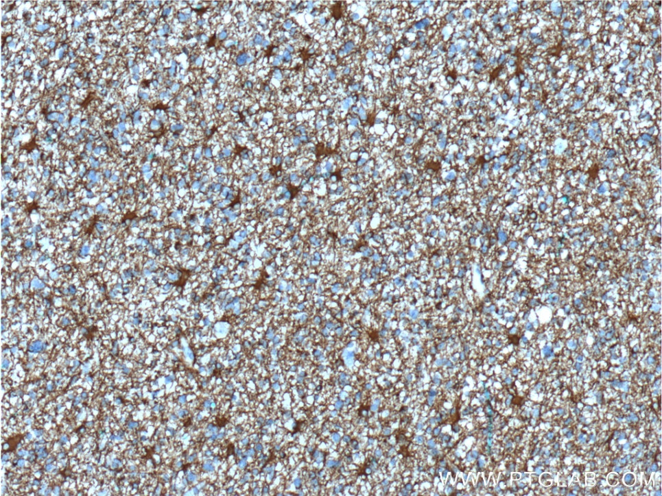
IHC staining of rat brain using 60190-1-Ig (same clone as 60190-1-PBS)
Immunohistochemical analysis of paraffin-embedded rat brain tissue slide using 60190-1-Ig (GFAP antibody) at dilution of 1:20000 (under 10x lens). Heat mediated antigen retrieval with Tris-EDTA buffer (pH 9.0). This data was developed using the same antibody clone with 60190-1-PBS in a different storage buffer formulation.
IHC staining of rat brain using 60190-1-Ig (same clone as 60190-1-PBS)
Immunohistochemical analysis of paraffin-embedded rat brain tissue slide using 60190-1-Ig (GFAP antibody) at dilution of 1:20000 (under 40x lens). Heat mediated antigen retrieval with Tris-EDTA buffer (pH 9.0). This data was developed using the same antibody clone with 60190-1-PBS in a different storage buffer formulation.
IHC staining of mouse brain using 60190-1-Ig (same clone as 60190-1-PBS)
Immunohistochemical analysis of paraffin-embedded mouse brain tissue slide using 60190-1-Ig (GFAP antibody) at dilution of 1:20000 (under 10x lens). Heat mediated antigen retrieval with Tris-EDTA buffer (pH 9.0). This data was developed using the same antibody clone with 60190-1-PBS in a different storage buffer formulation.
IHC staining of mouse brain using 60190-1-Ig (same clone as 60190-1-PBS)
Immunohistochemical analysis of paraffin-embedded mouse brain tissue slide using 60190-1-Ig (GFAP antibody) at dilution of 1:20000 (under 40x lens). Heat mediated antigen retrieval with Tris-EDTA buffer (pH 9.0). This data was developed using the same antibody clone with 60190-1-PBS in a different storage buffer formulation.
IHC staining of rat cerebellum using 60190-1-Ig (same clone as 60190-1-PBS)
Immunohistochemical analysis of paraffin-embedded rat cerebellum tissue slide using 60190-1-Ig (GFAP antibody) at dilution of 1:20000 (under 40x lens). Heat mediated antigen retrieval with Tris-EDTA buffer (pH 9.0). This data was developed using the same antibody clone with 60190-1-PBS in a different storage buffer formulation.
IHC staining of rat brain using 60190-1-Ig (same clone as 60190-1-PBS)
Immunohistochemical analysis of paraffin-embedded rat brain tissue slide using 60190-1-Ig (GFAP antibody) at dilution of 1:20000 (under 40x lens). Heat mediated antigen retrieval with Tris-EDTA buffer (pH 9.0). This data was developed using the same antibody clone with 60190-1-PBS in a different storage buffer formulation.
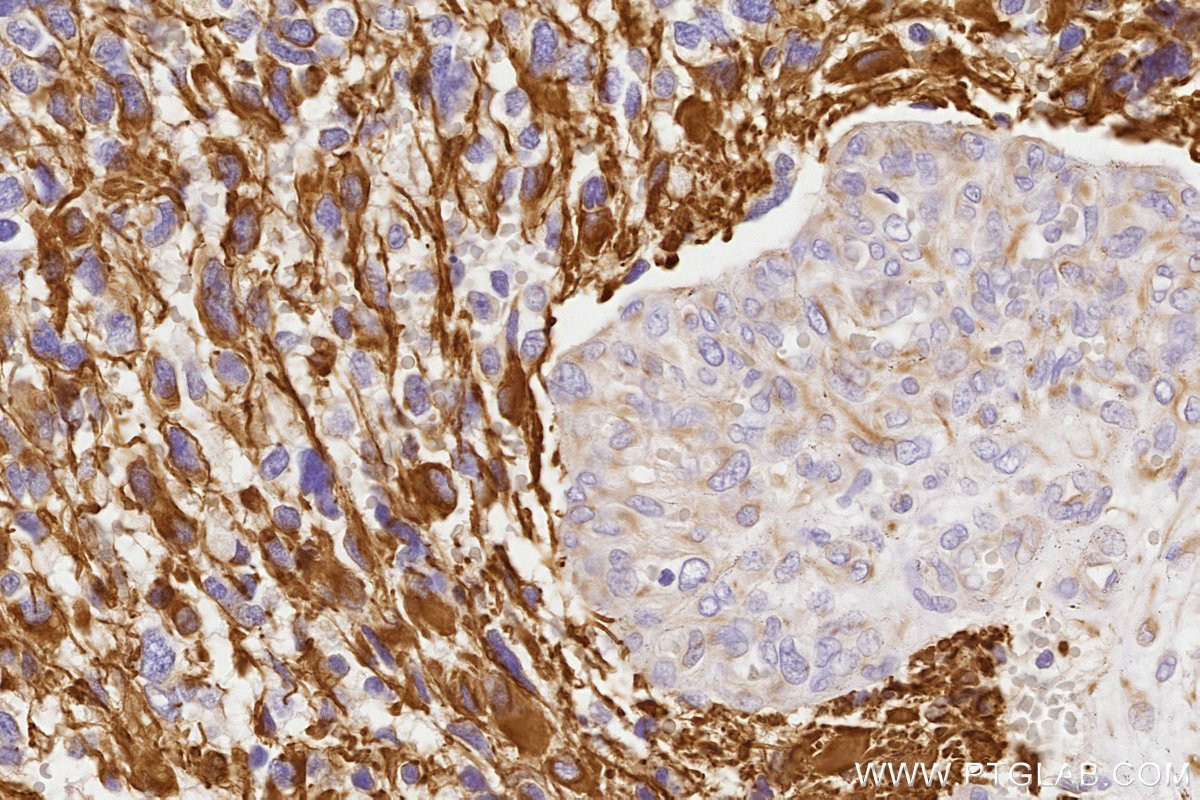
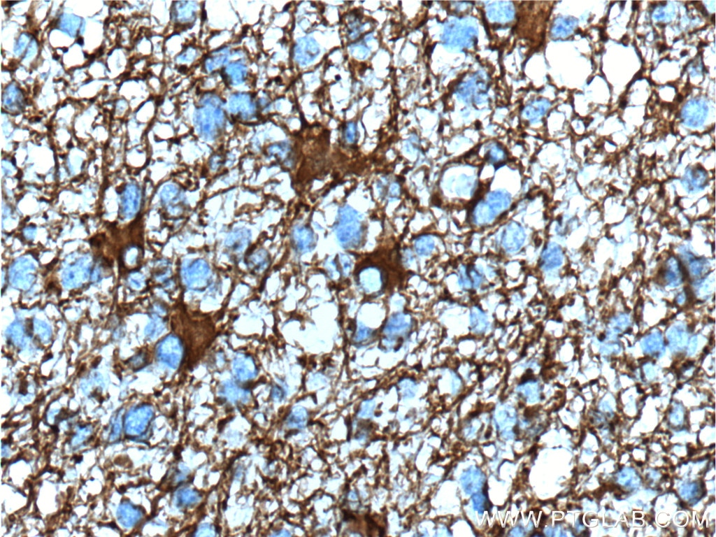
IHC staining of human gliomas using 60190-1-Ig (same clone as 60190-1-PBS)
Immunohistochemical analysis of paraffin-embedded human gliomas tissue slide using 60190-1-Ig (GFAP Antibody) at dilution of 1:5000 (under 10x lens). Heat mediated antigen retrieval with Tris-EDTA buffer (pH 9.0). This data was developed using the same antibody clone with 60190-1-PBS in a different storage buffer formulation.
IHC staining of human gliomas using 60190-1-Ig (same clone as 60190-1-PBS)
Immunohistochemical analysis of paraffin-embedded human gliomas tissue slide using 60190-1-Ig (GFAP Antibody) at dilution of 1:5000 (under 40x lens). Heat mediated antigen retrieval with Tris-EDTA buffer (pH 9.0). This data was developed using the same antibody clone with 60190-1-PBS in a different storage buffer formulation.
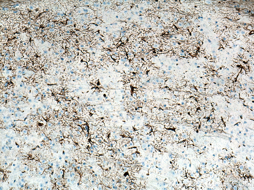
IHC staining of mouse brain using 60190-1-Ig (same clone as 60190-1-PBS)
Immunohistochemical analysis of paraffin-embedded mouse brain tissue slide using 60190-1-Ig (GFAP antibody) at dilution of 1:5000 (under 10x lens). Heat mediated antigen retrieval with Tris-EDTA buffer (pH 9.0). This data was developed using the same antibody clone with 60190-1-PBS in a different storage buffer formulation.
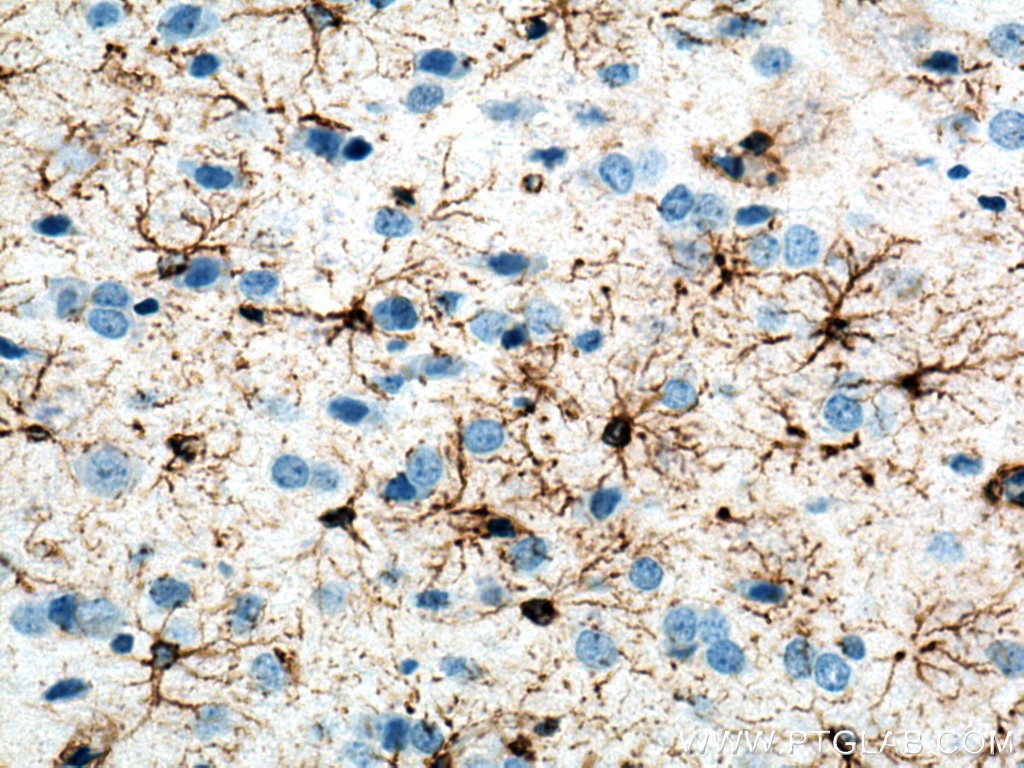
IHC staining of rat brain using 60190-1-Ig (same clone as 60190-1-PBS)
Immunohistochemical analysis of paraffin-embedded rat brain tissue slide using 60190-1-Ig (GFAP antibody) at dilution of 1:5000 (under 40x lens). Heat mediated antigen retrieval with Tris-EDTA buffer (pH 9.0). This data was developed using the same antibody clone with 60190-1-PBS in a different storage buffer formulation.
IF-P Figures
IF Staining of rat brain using 60190-1-Ig (same clone as 60190-1-PBS)
Immunofluorescent analysis of (4% PFA) fixed paraffin-embedded rat brain tissue using GFAP antibody (60190-1-Ig, Clone: 4B2E10 ) at dilution of 1:200 and CoraLite488-Conjugated AffiniPure Goat Anti-Mouse IgG(H+L). Heat mediated antigen retrieval with Tris-EDTA buffer (pH 9.0). This data was developed using the same antibody clone with 60190-1-PBS in a different storage buffer formulation.
IF Staining of mouse brain using 60190-1-Ig (same clone as 60190-1-PBS)
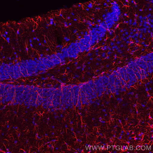
Immunofluorescent analysis of (4% PFA) fixed paraffin-embedded mouse brain tissue using GFAP antibody (60190-1-Ig, Clone: 4B2E10 ) at dilution of 1:800 and CoraLite®594-Conjugated AffiniPure Goat Anti-Mouse IgG(H+L). Heat mediated antigen retrieval with Tris-EDTA buffer (pH 9.0). This data was developed using the same antibody clone with 60190-1-PBS in a different storage buffer formulation.
IF Staining of rat brain using 60190-1-Ig (same clone as 60190-1-PBS)
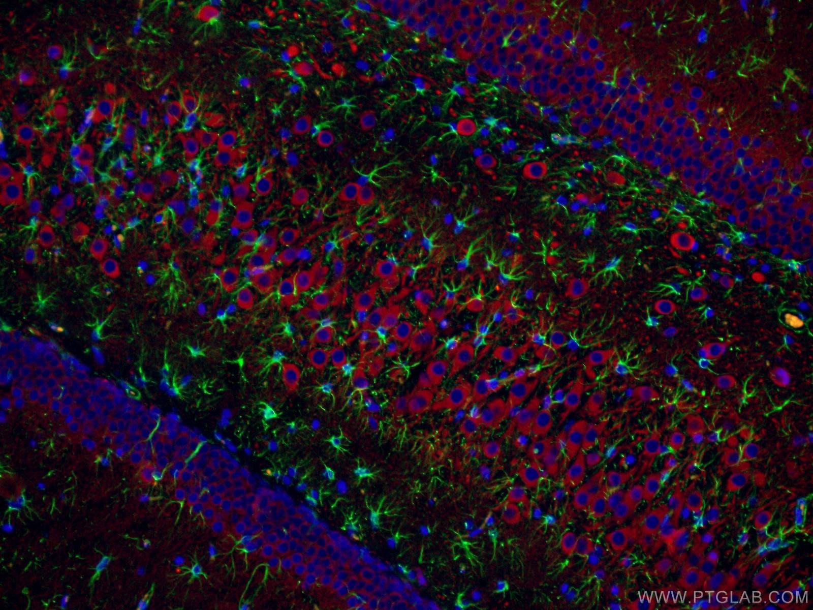
Immunofluorescent analysis of (4% PFA) fixed paraffin-embedded rat brain tissue using GFAP antibody (60190-1-Ig, Clone: 4B2E10 ) at dilution of 1:1000 and CoraLite®488-Conjugated Goat Anti-Mouse IgG(H+L) (SA00013-1), NeuN antibody (84401-4-RR, Clone: 241461G7, red). Heat mediated antigen retrieval with Tris-EDTA buffer (pH 9.0). This data was developed using the same antibody clone with 60190-1-PBS in a different storage buffer formulation.
IF Staining of rat brain using 60190-1-Ig (same clone as 60190-1-PBS)
Immunofluorescent analysis of (4% PFA) fixed paraffin-embedded rat brain tissue using GFAP antibody (60190-1-Ig, Clone: 4B2E10 ) at dilution of 1:1000 and CoraLite®488-Conjugated Goat Anti-Mouse IgG(H+L) (SA00013-1), NeuN antibody (84401-4-RR, Clone: 241461G7, red). Heat mediated antigen retrieval with Tris-EDTA buffer (pH 9.0). This data was developed using the same antibody clone with 60190-1-PBS in a different storage buffer formulation.
IF Staining of mouse brain using 60190-1-Ig (same clone as 60190-1-PBS)
Immunofluorescent analysis of (4% PFA) fixed paraffin-embedded mouse brain tissue using GFAP antibody (60190-1-Ig, Clone: 4B2E10 ) at dilution of 1:400 and CoraLite®594-Conjugated Goat Anti-Mouse IgG(H+L) (SA00013-3), S100B antibody (82271-8-RR, Clone: 1E2, green). Heat mediated antigen retrieval with Tris-EDTA buffer (pH 9.0). This data was developed using the same antibody clone with 60190-1-PBS in a different storage buffer formulation.
IF Staining of mouse brain using 60190-1-Ig (same clone as 60190-1-PBS)
Immunofluorescent analysis of (4% PFA) fixed paraffin-embedded mouse brain tissue using GFAP antibody (60190-1-Ig, Clone: 4B2E10 ) at dilution of 1:1500 and Rhodamine (TRITC)-conjugated Goat Anti-Mouse IgG(H+L) (SA00007-1), TMEM119 antibody (83448-9-RR, Clone: 251557E2, green), CoraLite® Plus 647 IBA1 antibody (CL647-81728, Clone: 4D5, Magenta). Heat mediated antigen retrieval with Tris-EDTA buffer (pH 9.0). This data was developed using the same antibody clone with 60190-1-PBS in a different storage buffer formulation.