
Validation Data Gallery
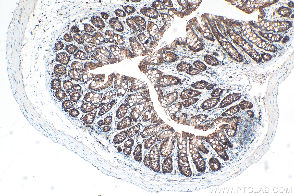
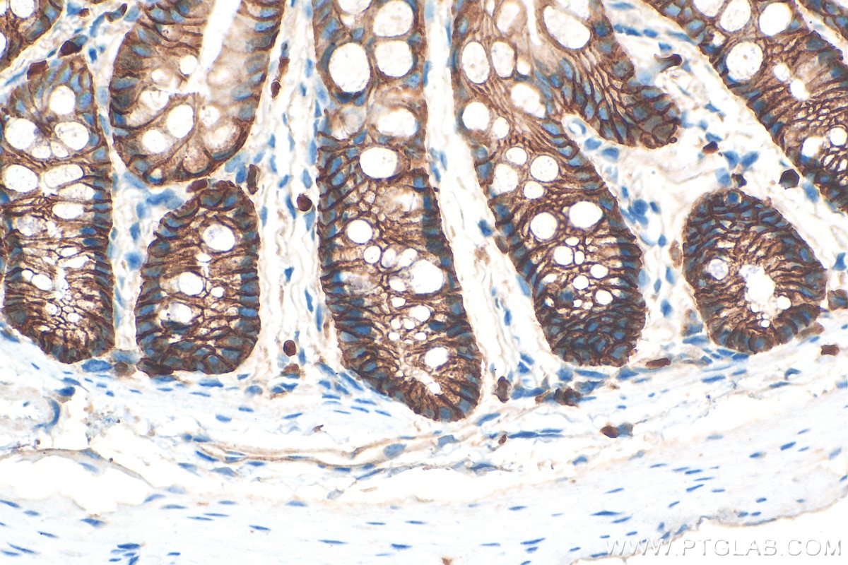
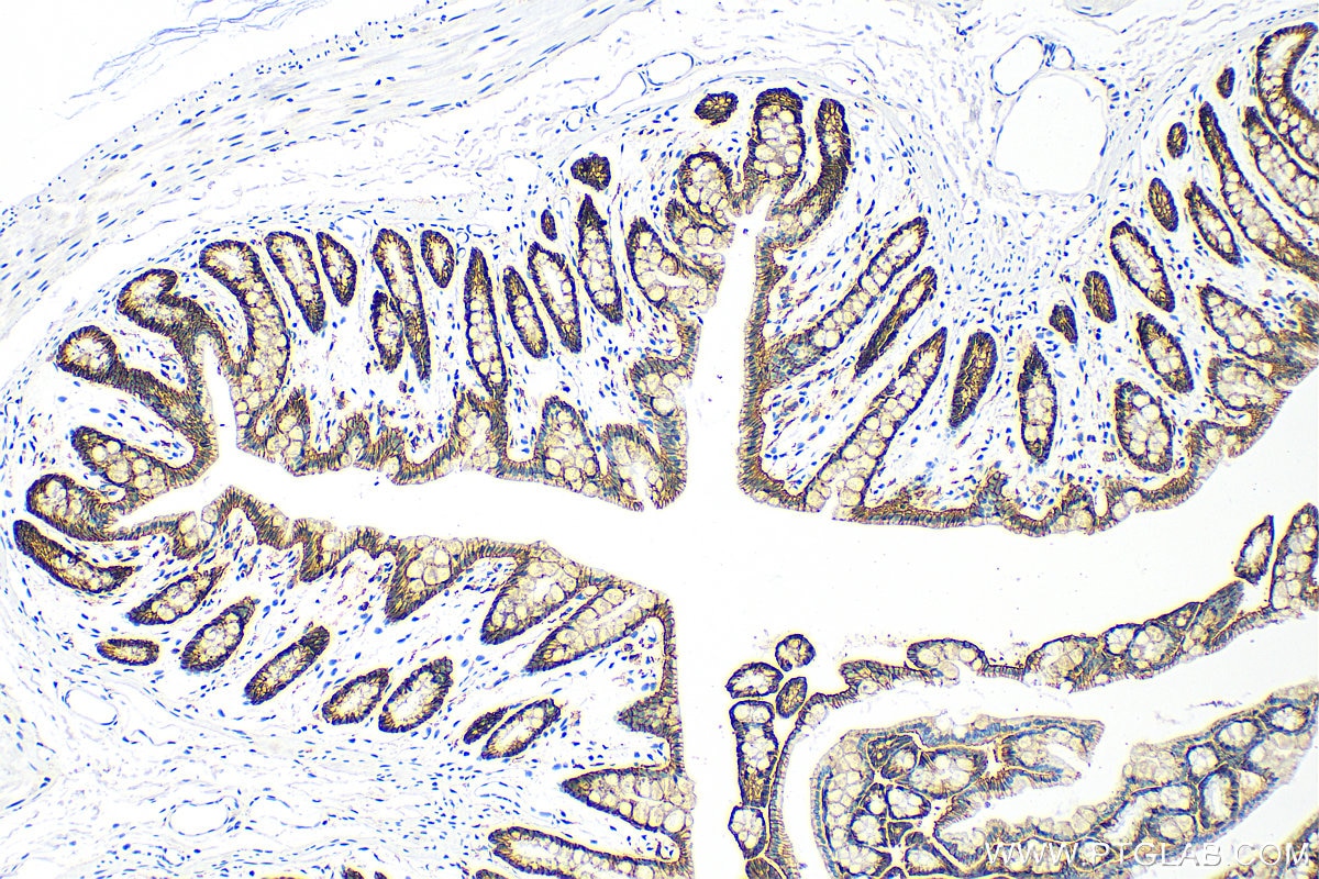
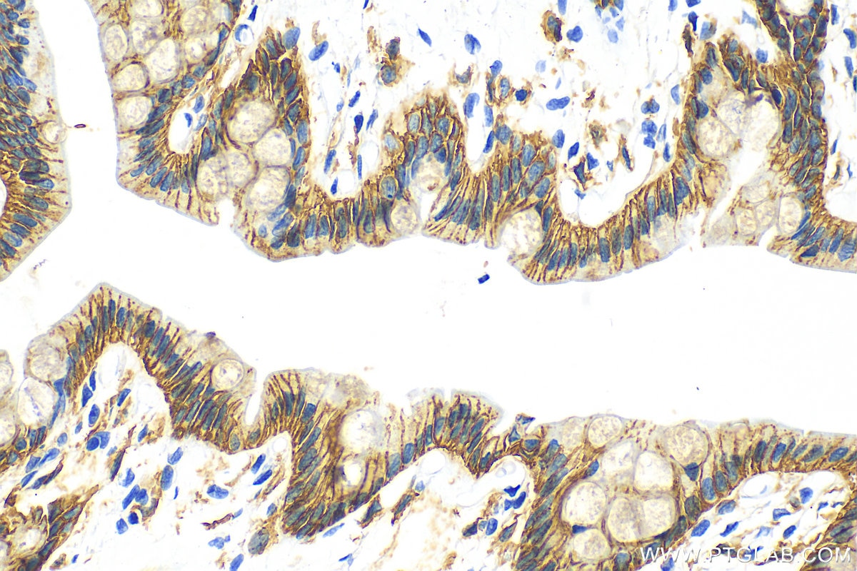
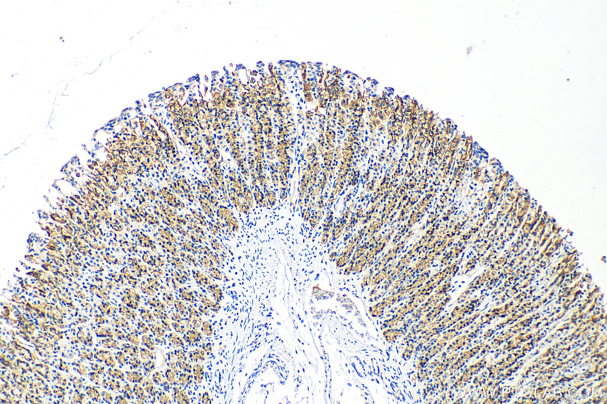
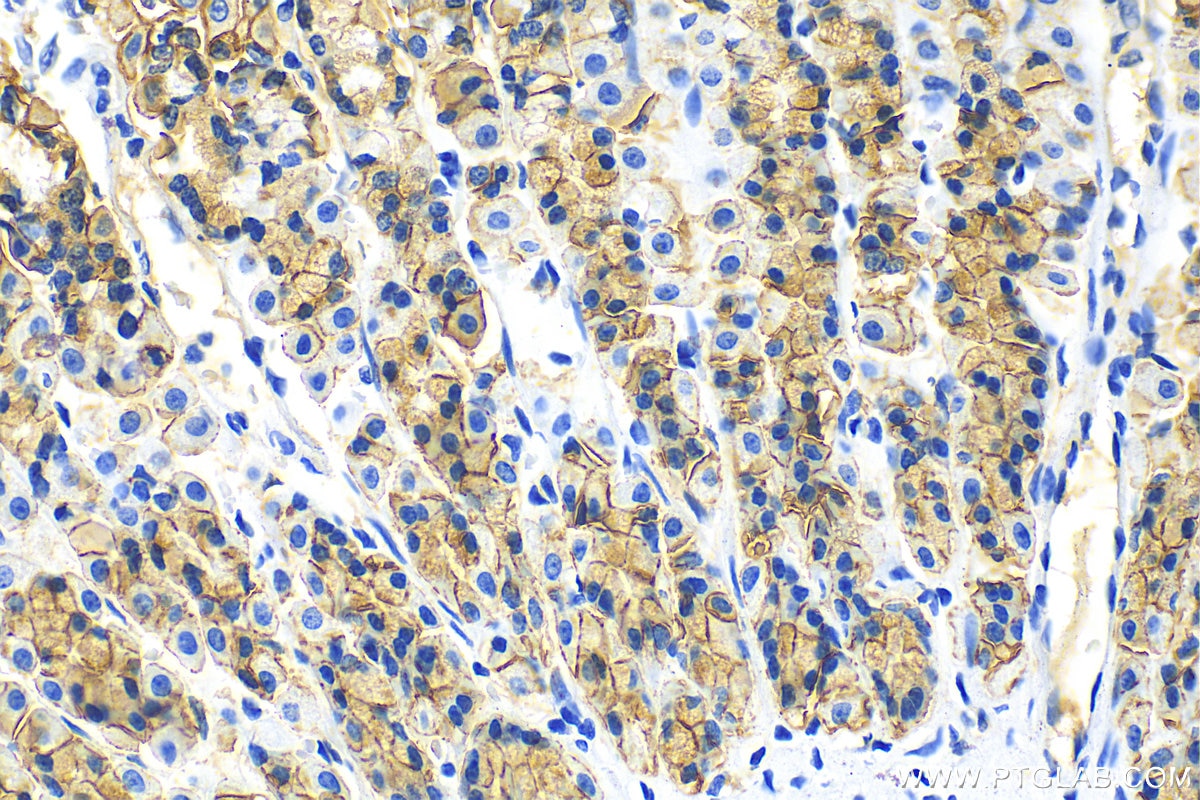
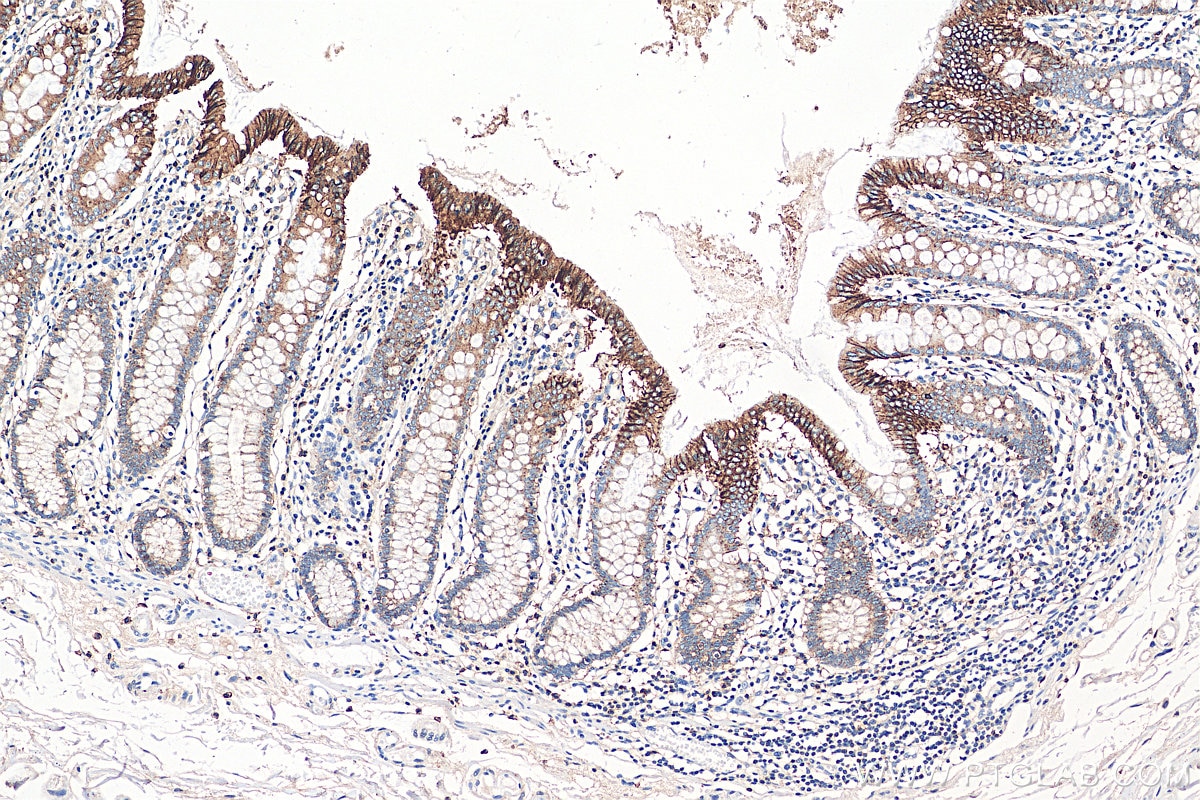
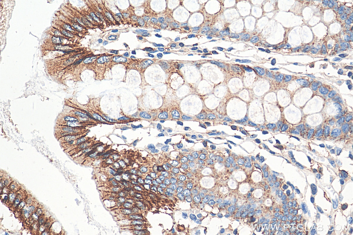
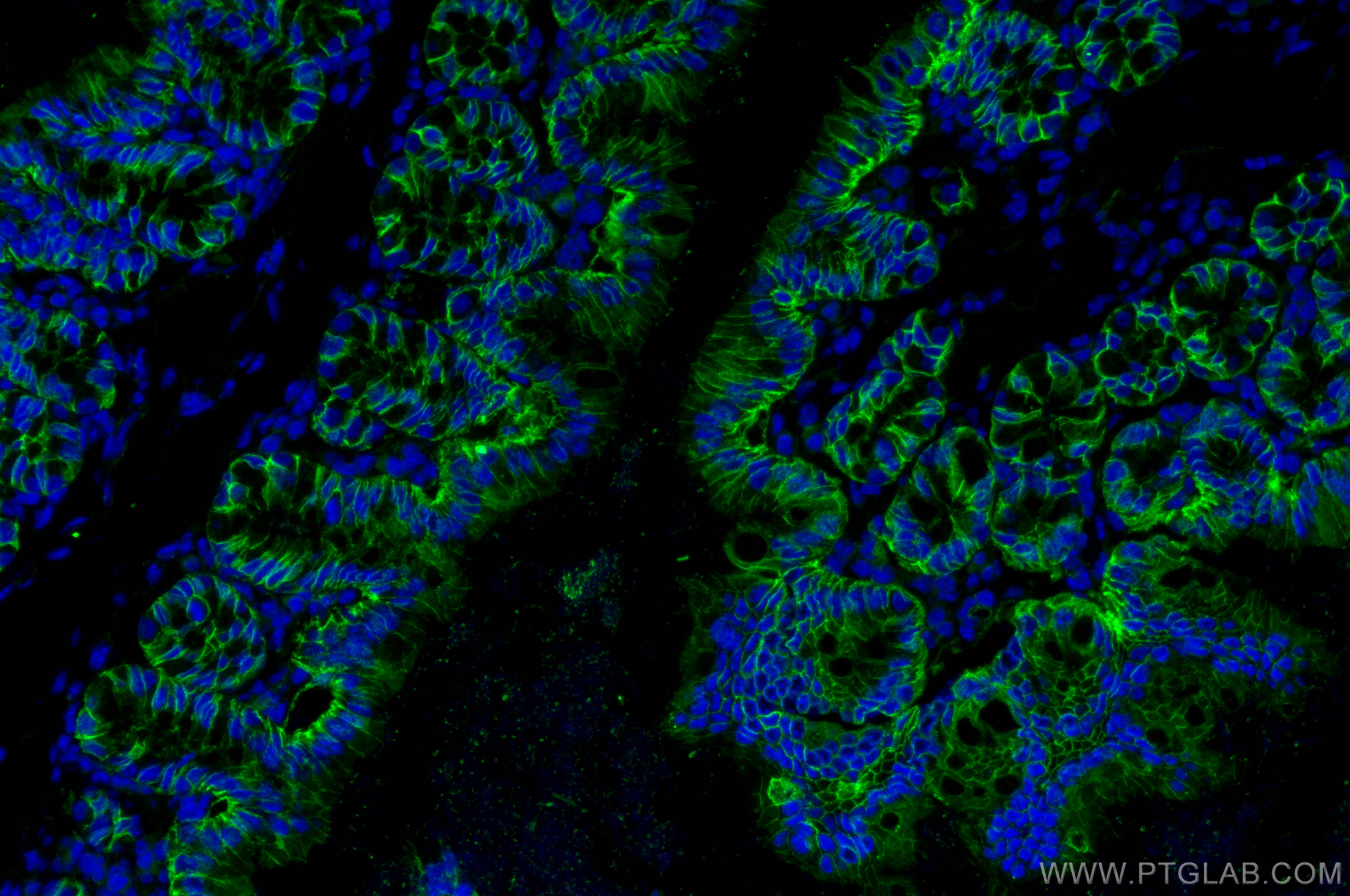
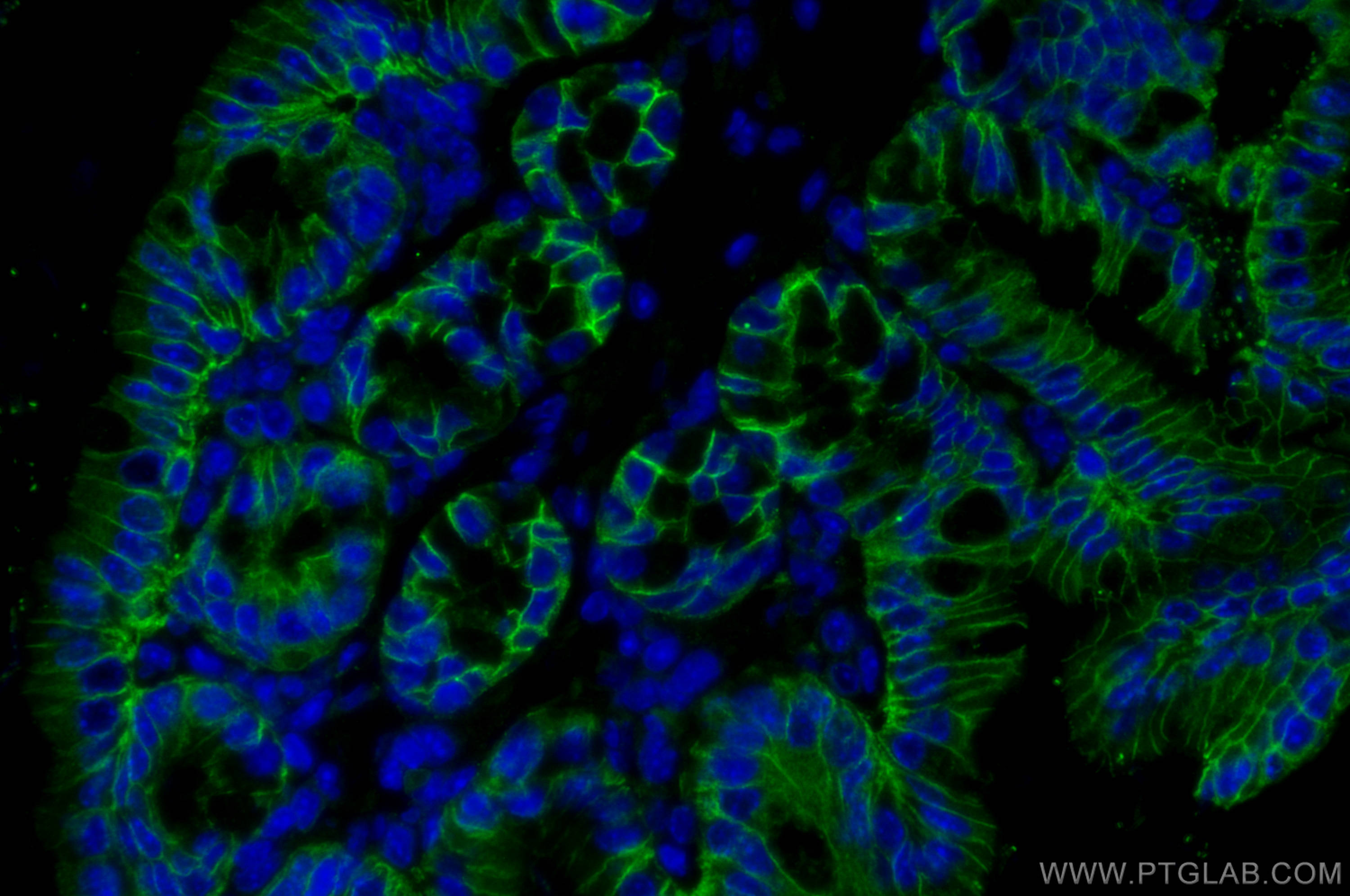
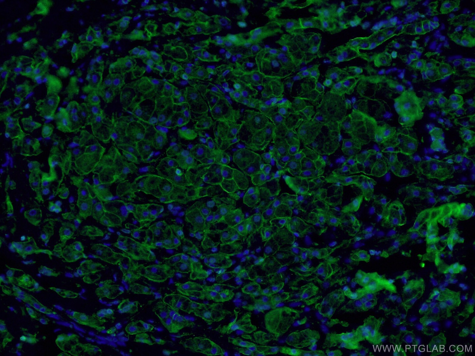
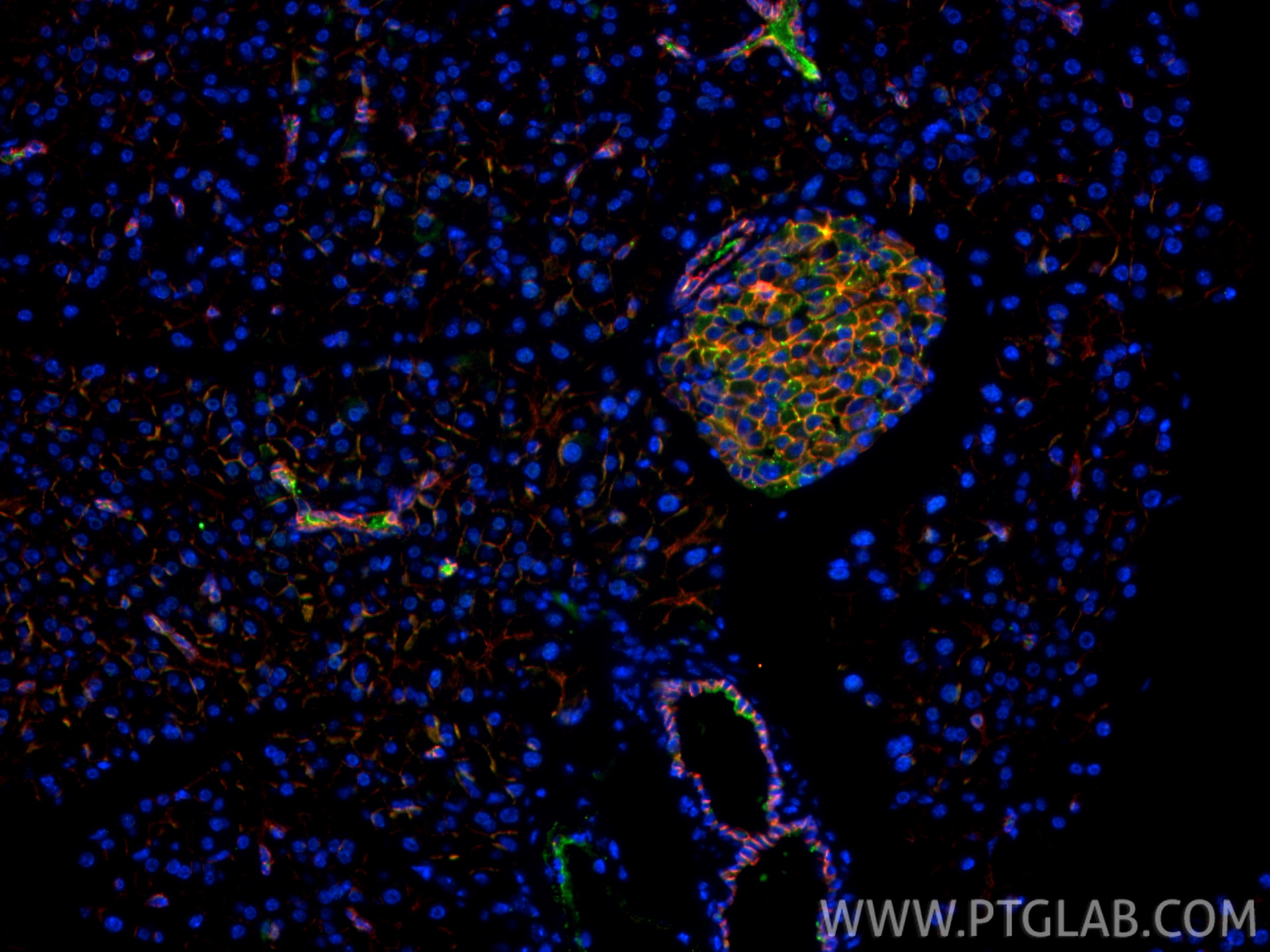
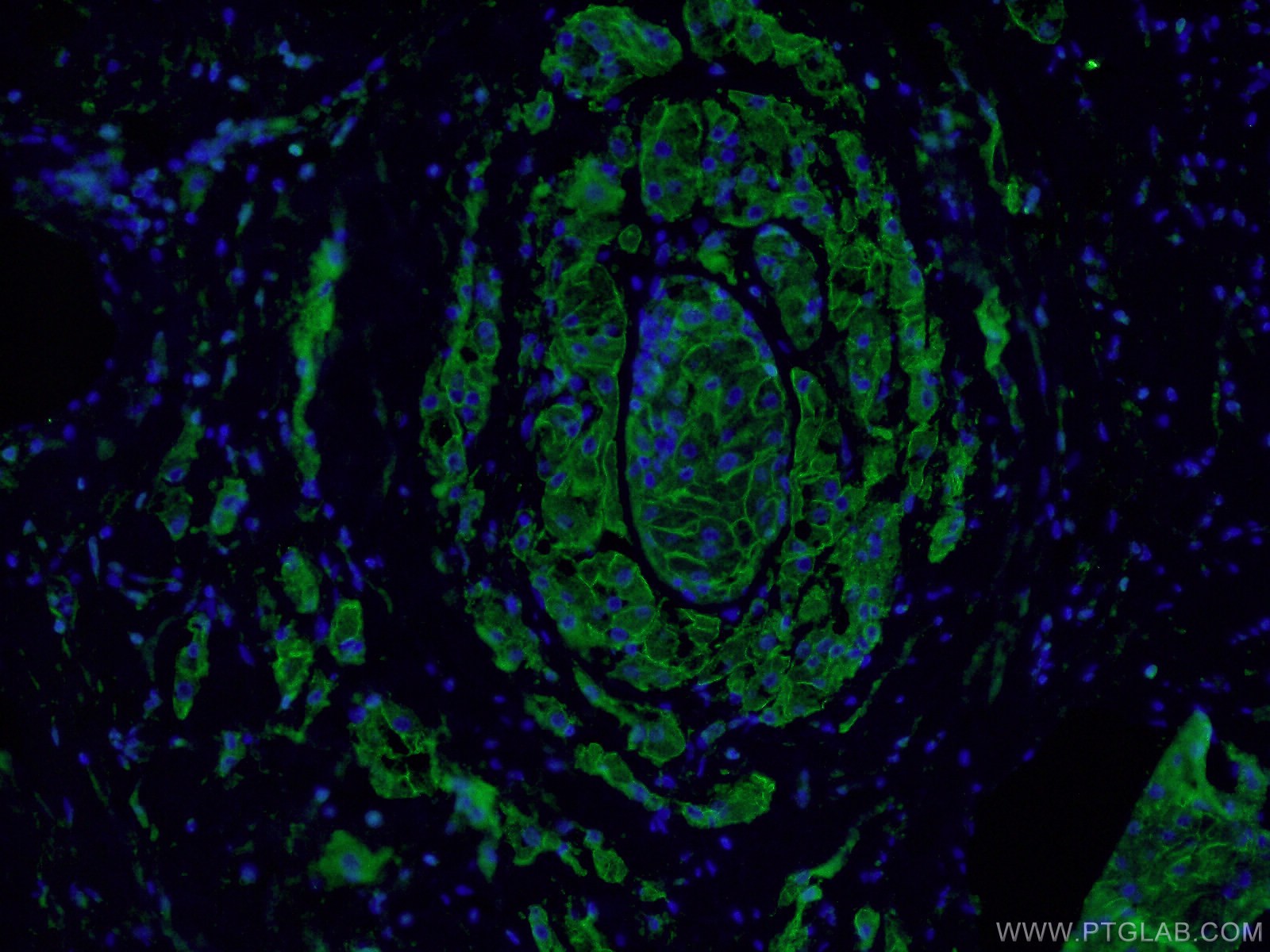
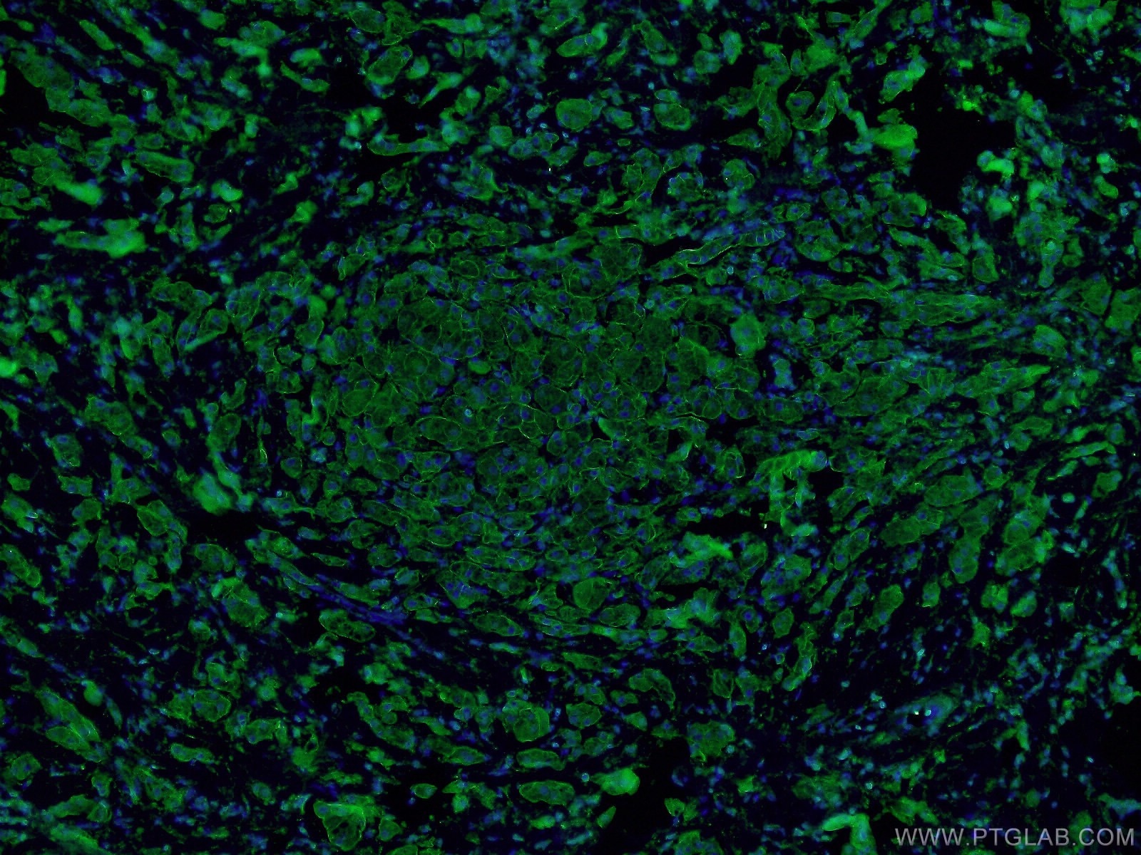
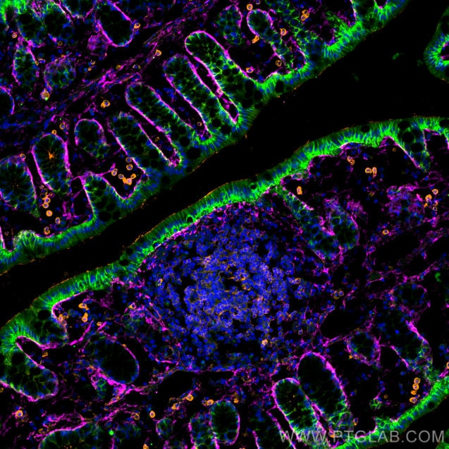
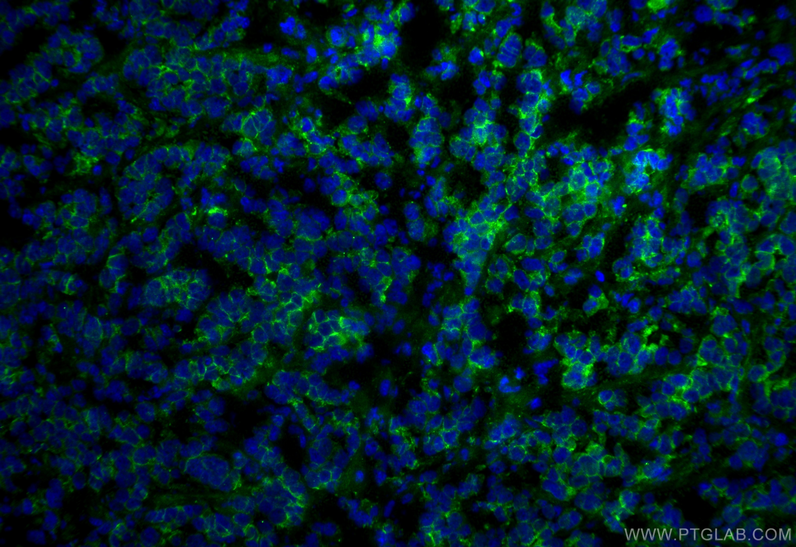
Tested Applications
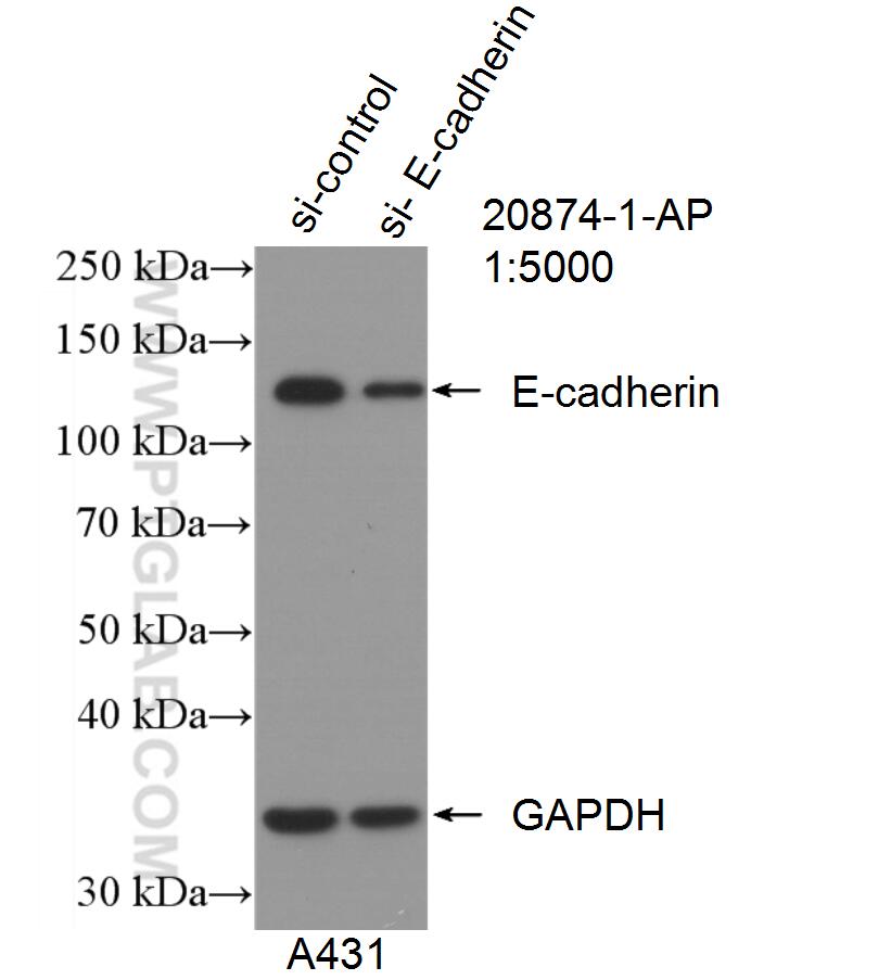
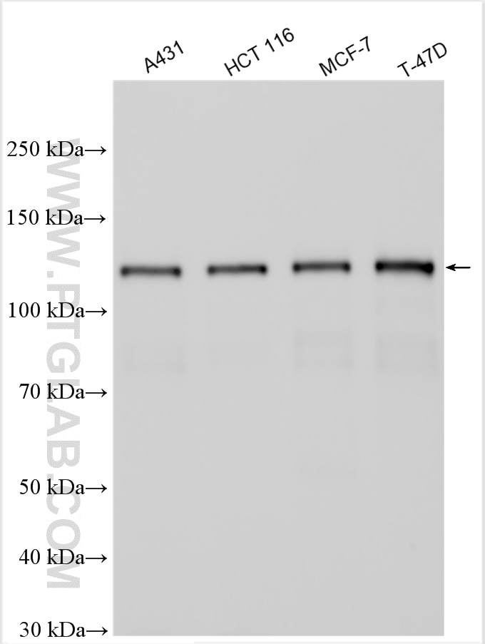
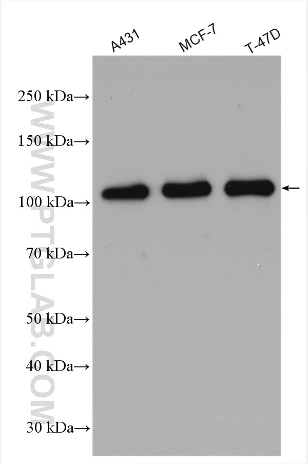
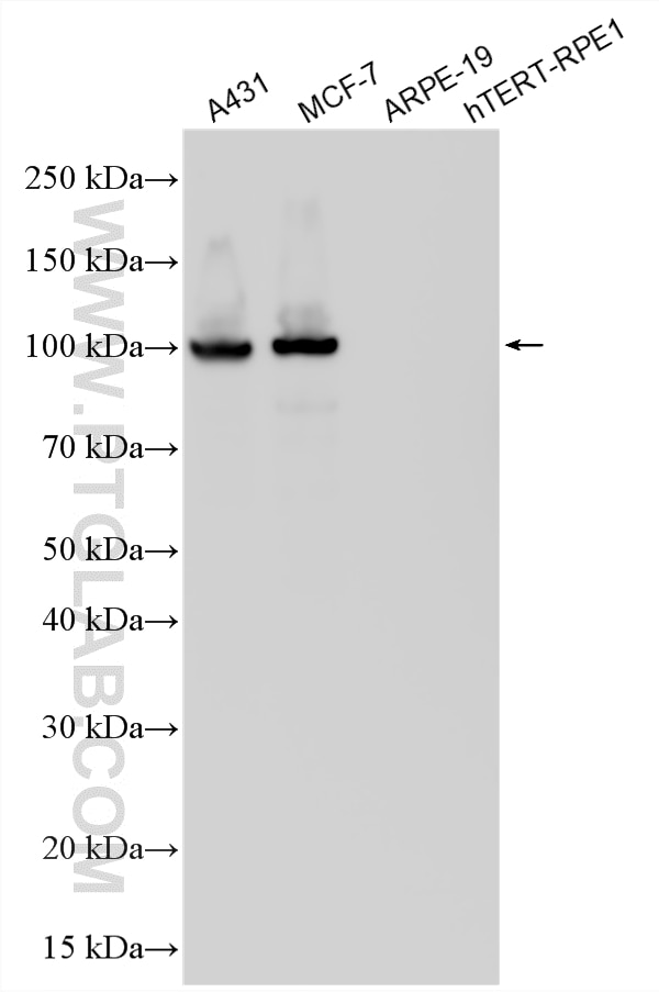
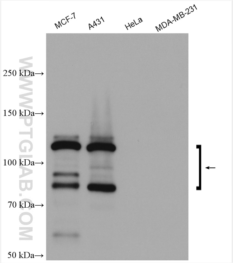
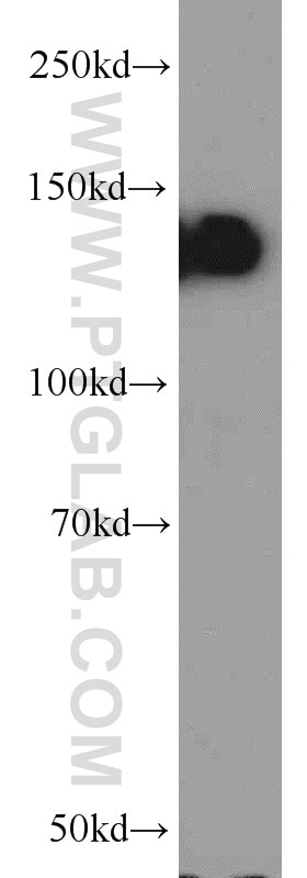
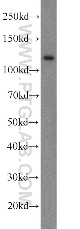
| Positive WB detected in | A431 cells, DU 145 cells, mouse testis tissue, HCT 116 cells, MCF-7 cells, T-47D cells |
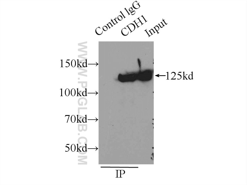
| Positive IP detected in | A431 cells |
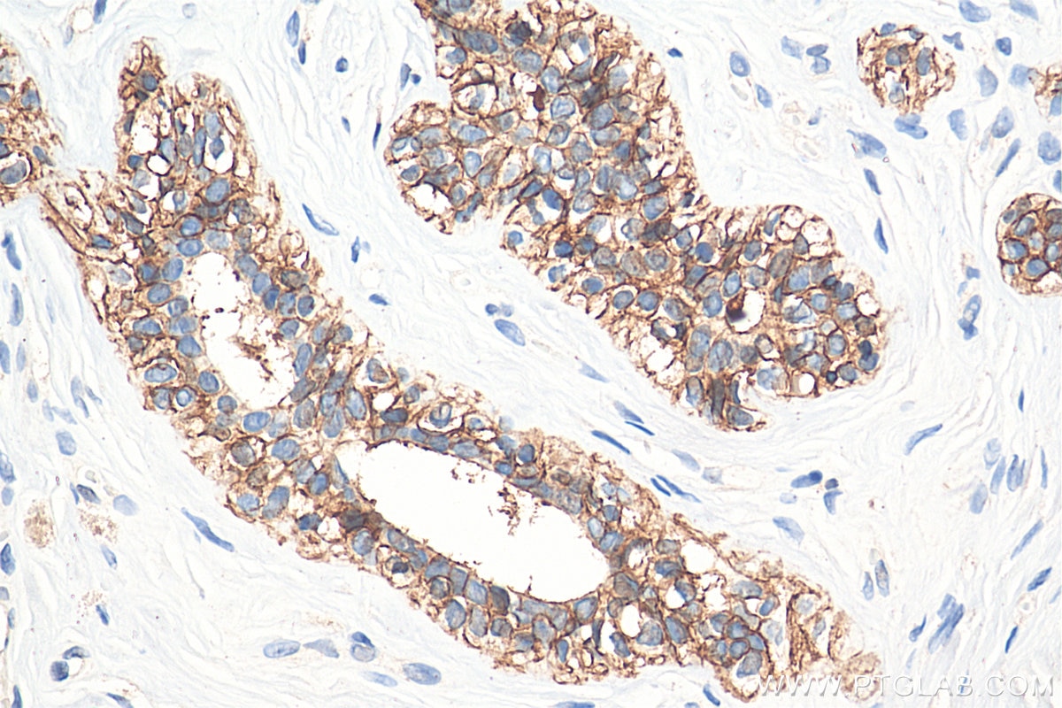
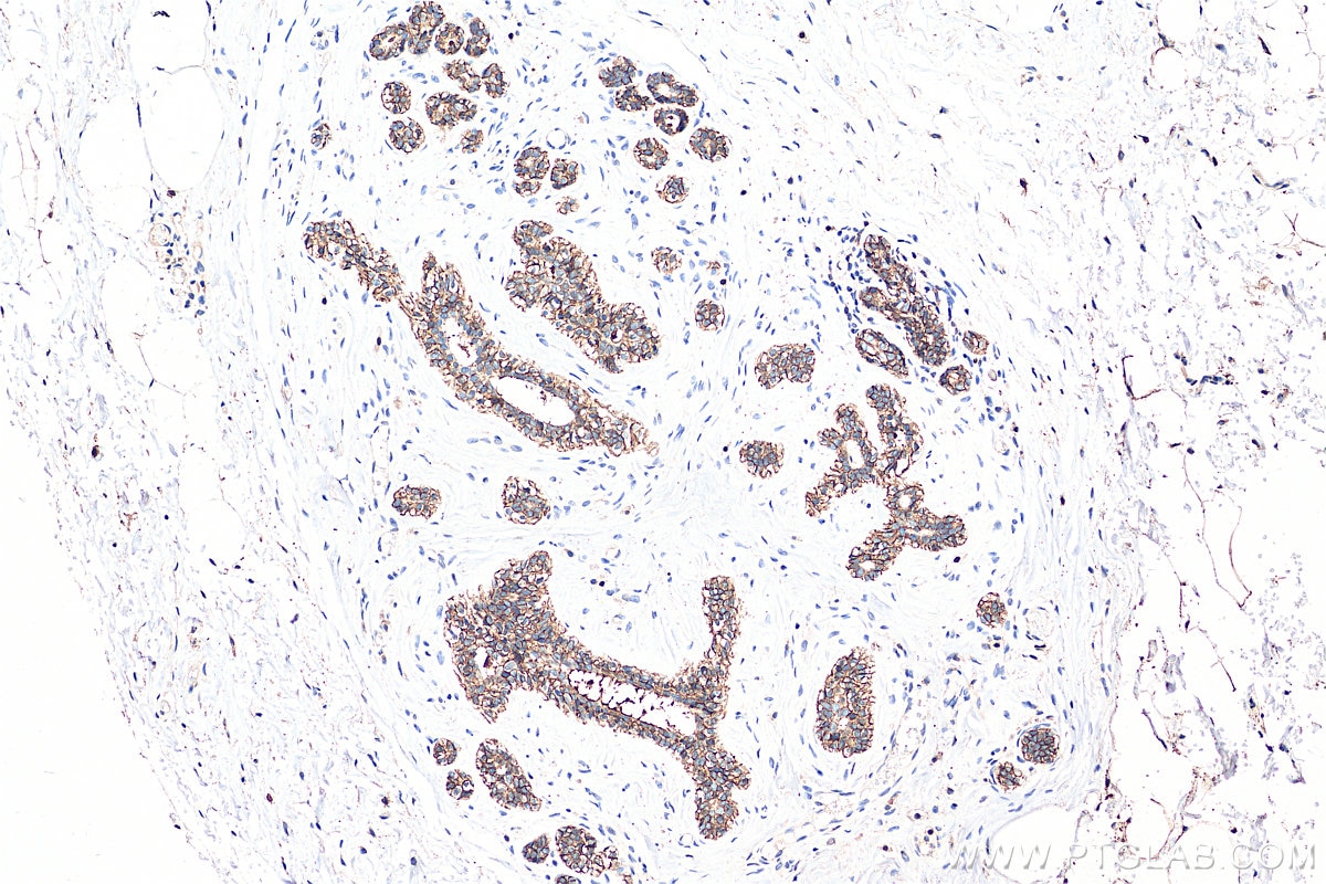
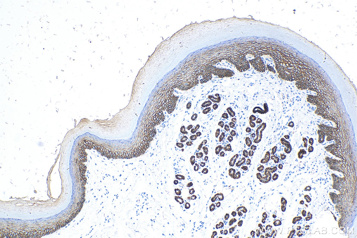
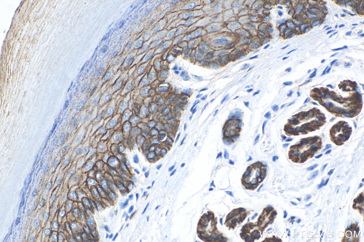
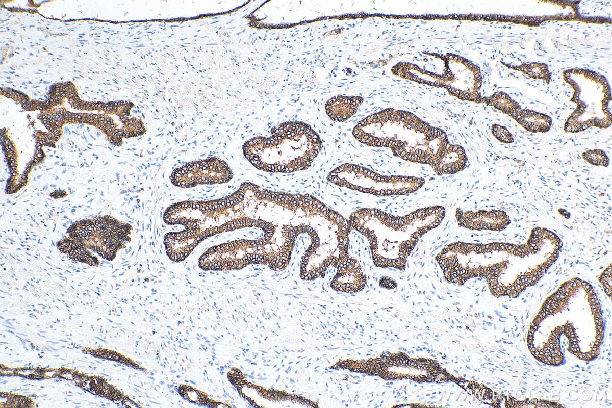
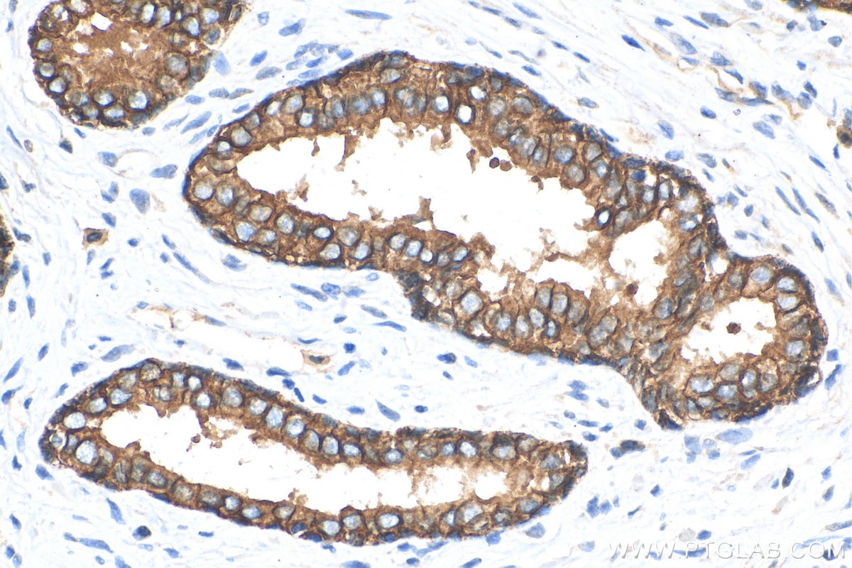
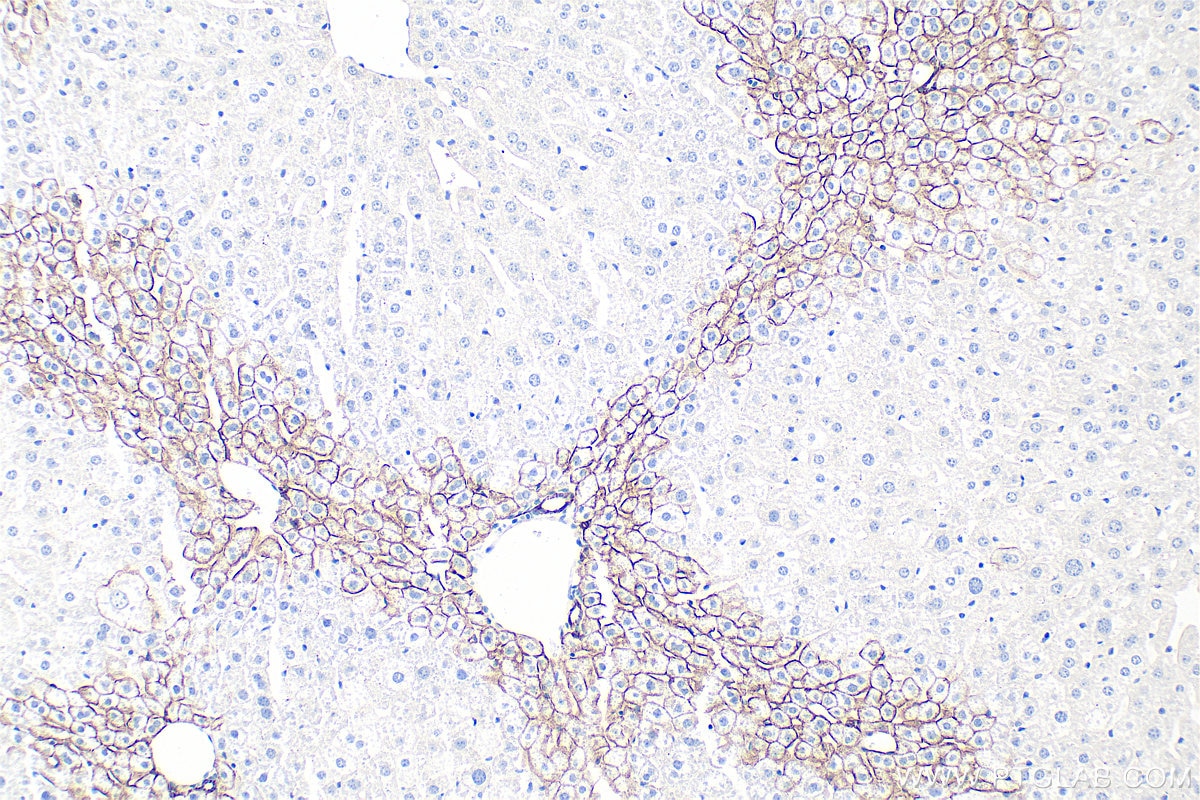
| Positive IHC detected in | mouse colon tissue, human breast cancer tissue, human colon tissue, human prostate cancer tissue, mouse liver tissue, mouse skin tissue, rat colon tissue, rat stomach tissue Note: suggested antigen retrieval with TE buffer pH 9.0; (*) Alternatively, antigen retrieval may be performed with citrate buffer pH 6.0 |
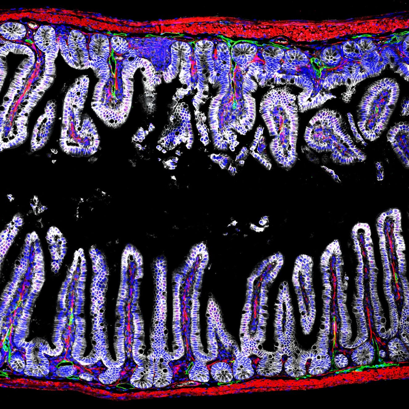
| Positive IF-P detected in | mouse colon tissue, human breast cancer tissue, mouse small intestine tissue, mouse pancreas tissue |
| Positive IF-Fro detected in | mouse colon tissue, mouse breast cancer |
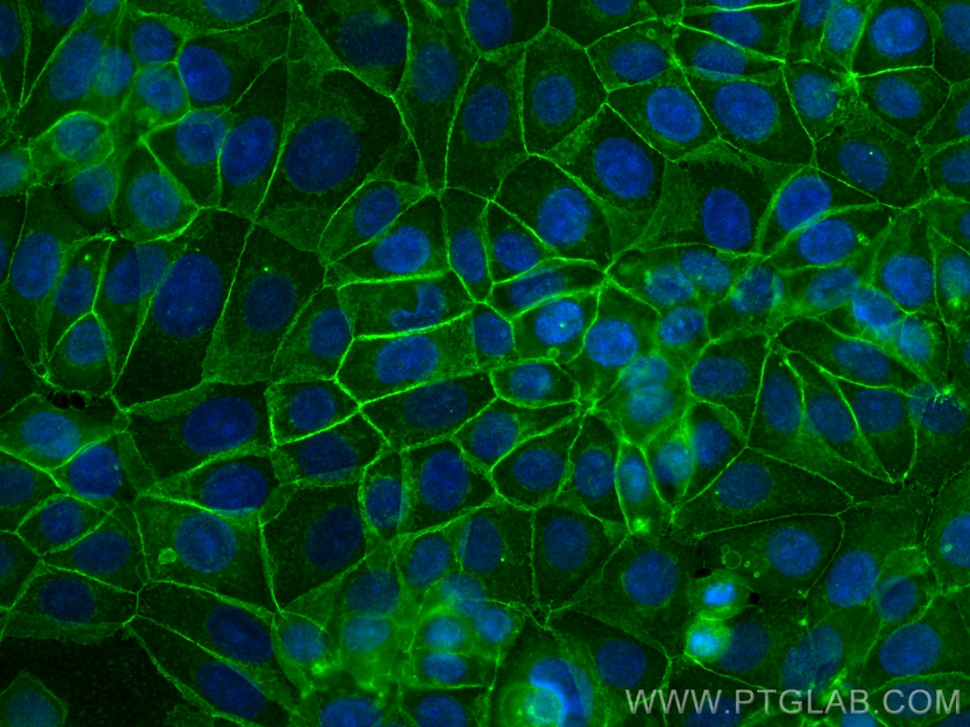
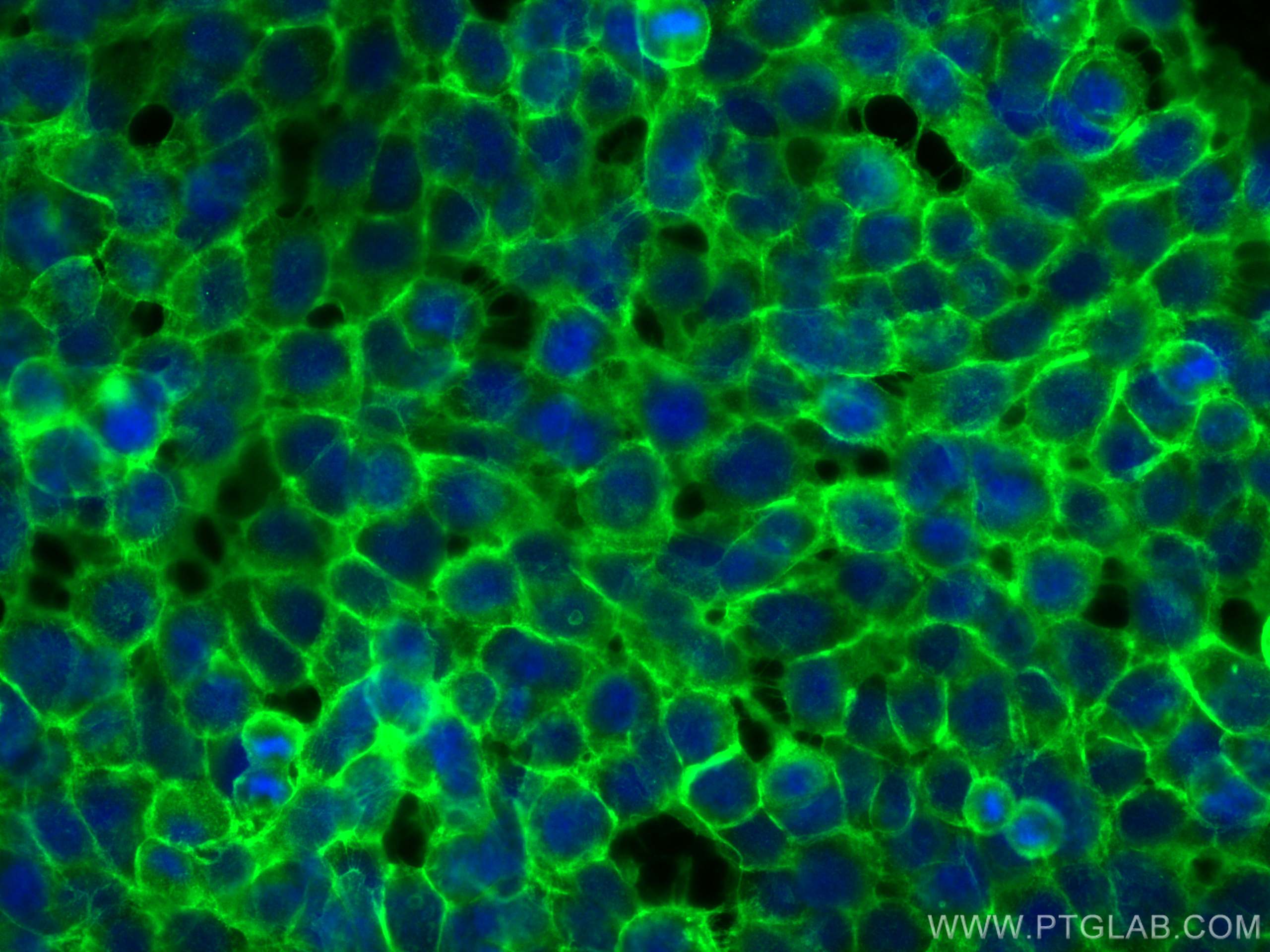
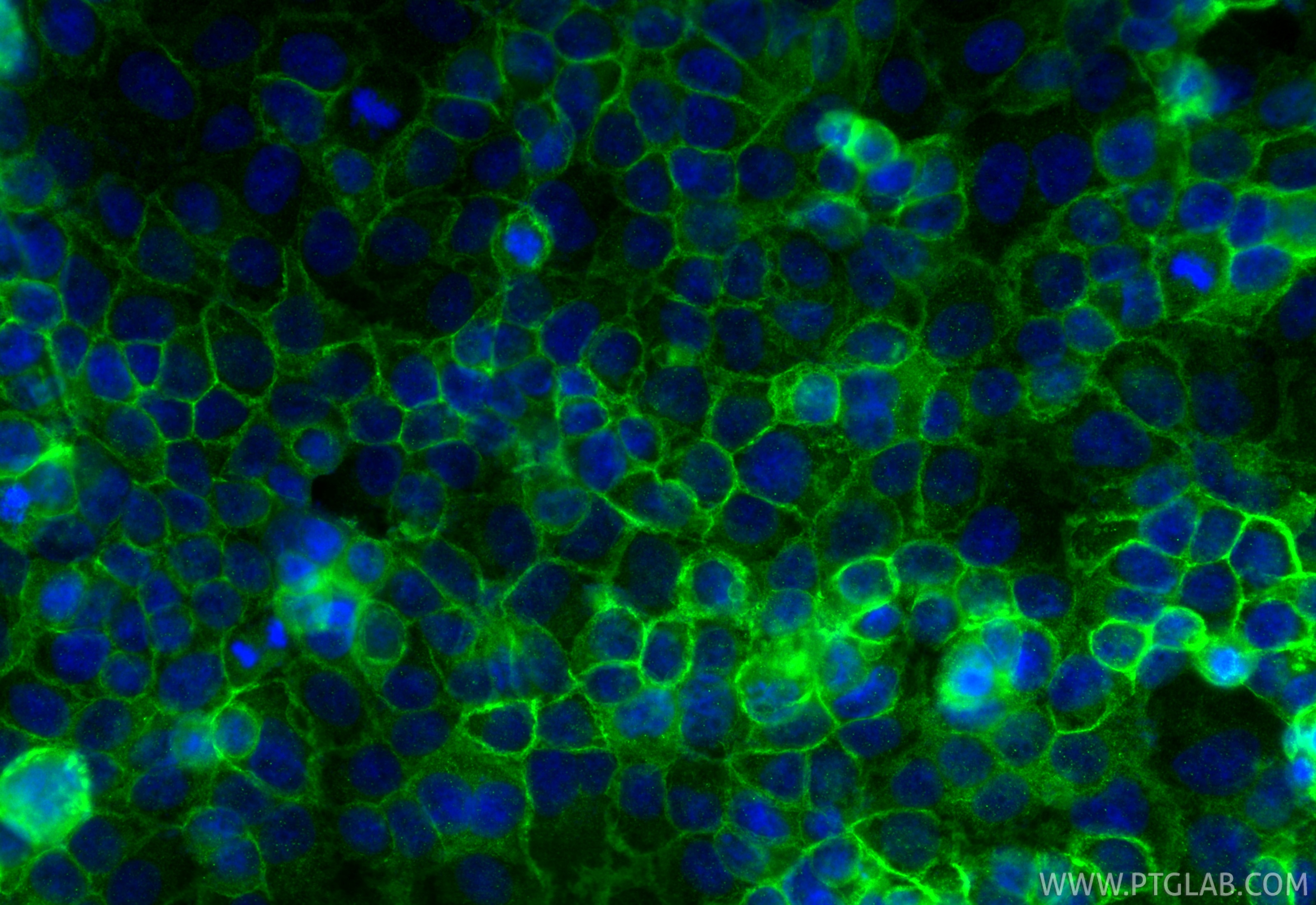
| Positive IF/ICC detected in | MCF-7 cells, A431 cells, OVCAR-3 cells |
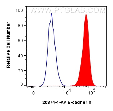
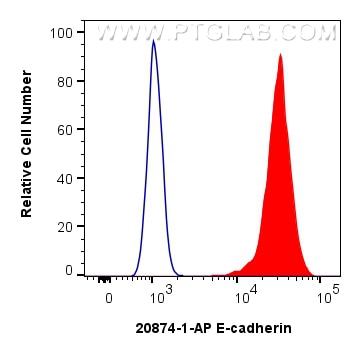
| Positive FC (Intra) detected in | MCF-7 cells |
| Positive FC detected in | MCF-7 cells |
Some bands between 80 and 120 kDa may be observed due to proteolytic cleavage.
Recommended dilution
| Application | Dilution |
|---|---|
| Western Blot (WB) | WB : 1:20000-1:200000 |
| Immunoprecipitation (IP) | IP : 0.5-4.0 ug for 1.0-3.0 mg of total protein lysate |
| Immunohistochemistry (IHC) | IHC : 1:5000-1:20000 |
| Immunofluorescence (IF)-P | IF-P : 1:200-1:800 |
| Immunofluorescence (IF)-FRO | IF-FRO : 1:50-1:500 |
| Immunofluorescence (IF)/ICC | IF/ICC : 1:200-1:800 |
| Flow Cytometry (FC) (INTRA) | FC (INTRA) : 0.25 ug per 10^6 cells in a 100 µl suspension |
| Flow Cytometry (FC) | FC : 0.25 ug per 10^6 cells in a 100 µl suspension |
| It is recommended that this reagent should be titrated in each testing system to obtain optimal results. | |
| Sample-dependent, Check data in validation data gallery. | |
Product Information
20874-1-AP targets E-cadherin in WB, IHC, IF/ICC, IF-P, IF-Fro, FC (Intra), IP, CoIP, ELISA applications and shows reactivity with human, mouse, rat samples.
| Tested Reactivity | human, mouse, rat |
| Cited Reactivity | human, mouse, rat, pig, canine, chicken, zebrafish, bovine |
| Host / Isotype | Rabbit / IgG |
| Class | Polyclonal |
| Type | Antibody |
| Immunogen |
CatNo: Ag14973 Product name: Recombinant human E-cadherin protein Source: e coli.-derived, PGEX-4T Tag: GST Domain: 373-622 aa of BC141838 Sequence: PIFNPTTYKGQVPENEANVVITTLKVTDADAPNTPAWEAVYTILNDDGGQFVVTTNPVNNDGILKTAKGLDFEAKQQYILHVAVTNVVPFEVSLTTSTATVTVDVLDVNEAPIFVPPEKRVEVSEDFGVGQEITSYTAQEPDTFMEQKITYRIWRDTANWLEINPDTGAISTRAELDREDFEHVKNSTYTALIIATDNGSPVATGTGTLLLILSDVNDNAPIPEPRTIFFCERNPKPQVINIIDADLPPI 相同性解析による交差性が予測される生物種 |
| Full Name | cadherin 1, type 1, E-cadherin (epithelial) |
| Calculated molecular weight | 882 aa, 97 kDa |
| Observed molecular weight | 120-125 kDa |
| GenBank accession number | BC141838 |
| Gene Symbol | E-cadherin |
| Gene ID (NCBI) | 999 |
| RRID | AB_10697811 |
| Conjugate | Unconjugated |
| Form | |
| Form | Liquid |
| Purification Method | Antigen affinity purification |
| UNIPROT ID | P12830 |
| Storage Buffer | PBS with 0.02% sodium azide and 50% glycerol{{ptg:BufferTemp}}7.3 |
| Storage Conditions | Store at -20°C. Stable for one year after shipment. Aliquoting is unnecessary for -20oC storage. |
Background Information
Cadherins are a family of transmembrane glycoproteins that mediate calcium-dependent cell-cell adhesion and play an important role in the maintenance of normal tissue architecture. E-cadherin (epithelial cadherin), also known as CDH1 (cadherin 1) or CAM 120/80, is a classical member of the cadherin superfamily which also includes N-, P-, R-, and B-cadherins. E-cadherin is expressed on the cell surface in most epithelial tissues. The extracellular region of E-cadherin establishes calcium-dependent homophilic trans binding, providing specific interaction with adjacent cells, while the cytoplasmic domain is connected to the actin cytoskeleton through the interaction with p120-, α-, β-, and γ-catenin (plakoglobin). E-cadherin is important in the maintenance of epithelial integrity and is involved in mechanisms regulating proliferation, differentiation, and survival of epithelial cells. E-cadherin may also play a role in tumorigenesis. It is considered to be an invasion suppressor protein, and its loss is an indicator of high tumor aggressiveness. E-cadherin is sensitive to trypsin digestion in the absence of Ca2+. This polyclonal antibody recognizes 120-125 kDa intact E-cadherin and its cleaved fragments of 80-120 kDa.
Protocols
| Product Specific Protocols | |
|---|---|
| FC protocol for E-cadherin antibody 20874-1-AP | Download protocol |
| IF protocol for E-cadherin antibody 20874-1-AP | Download protocol |
| IHC protocol for E-cadherin antibody 20874-1-AP | Download protocol |
| IP protocol for E-cadherin antibody 20874-1-AP | Download protocol |
| WB protocol for E-cadherin antibody 20874-1-AP | Download protocol |
| Standard Protocols | |
|---|---|
| Click here to view our Standard Protocols |
Publications
| Species | Application | Title |
|---|---|---|
Signal Transduct Target Ther Prostaglandin E2-driven dedifferentiation of Schwann cells leads to perineural invasion in pancreatic ductal adenocarcinoma | ||
Signal Transduct Target Ther Hypoxia-inducible factor upregulation by roxadustat attenuates drug reward by altering brain iron homoeostasis | ||
Cancer Cell A conserved re-epithelialization program underlies malignancy in pancreatic ductal adenocarcinoma. | ||
J Hematol Oncol LincK contributes to breast tumorigenesis by promoting proliferation and epithelial-to-mesenchymal transition. | ||
J Hematol Oncol The LINC00623/NAT10 signaling axis promotes pancreatic cancer progression by remodeling ac4C modification of mRNA |