Validation Data Gallery
Product Information
DNA plasmid encoding for anti-PCNA VHH (anti-PCNA Nanobody) fused to fluorescent protein TagRFP. PCNA is a cell cycle marker.
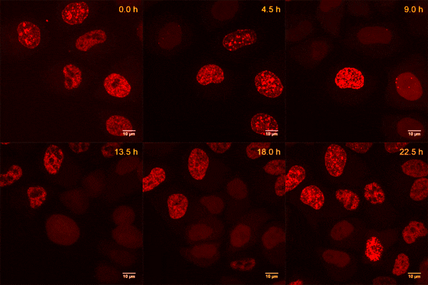
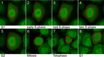
| Description | The Cell Cycle-Chromobody allows visualization of the endogenous cell cycle marker protein PCNA: • Trace dynamic changes during the cell cycle in real-time • Monitor the distribution of an endogenous cell cycle marker protein without artifacts or cytotoxic effects when overexpressing fluorescent fusion proteins • Does not affect cellular functions
|
| Applications | Live cell imaging Visualizing endogenous cell cycle marker protein PCNA Actin as biomarker or control |
| Specificity | PCNA (proliferating cell nuclear antigen), tested in human cells and zebrafish |
| Reporter | TagRFP |
| Vector type | Mammalian expression vector |
| Promoter | Constitutive CMV IE |
| Codon usage | Mammalian |
| Selection | Kan/Neo |
| Sequence | With the Cell Cycle-Chromobody plasmid you receive the sequence information of the Alpaca antibody to PCNA fused to TagRFP, as well as the full vector sequence. |
| Transfection | Transfection of Chromobody plasmids into mammalian cells can be done with standard DNA-transfection methods, e.g. lipofection (Lipofectamine 2000® from Thermo Scientific), according to the manufacturer’s protocol for the transfection reagent. Please choose the transfection method that works the best for your cell type. |
| Microscopy techniques | Wide-field epifluorescence microscopy; confocal microscopy; super-resolution microscopy e.g. STED and 3D-SIM; high-content microscopy and analysis |
| Storage Condition | Shipped at ambient temperature. Store at -20 °C |
Documentation
| SDS |
|---|
| ccr_SDS_Cell Cycle Chromobody® plasmid (TagRFP) (EN) |
| Datasheet |
|---|
| Cell Cycle Chromobody® plasmid (TagRFP) Datasheet |
| EULA |
|---|
| ccr End User License Agreement (EULA) Cell Cycle-Chromobody TagRFP plasmid non-profit (PDF) |
| ccr End User License Agreement (EULA) Cell Cycle-Chromobody TagRFP plasmid non-profit (US) (PDF) |
| Brochure |
|---|
| Chromotek nanobodies brochure (PDF) |
Publications
| Application | Title |
|---|---|
Nature Equilibrium between nascent and parental MCM proteins protects replicating genomes. | |
Nat Cell Biol 53BP1 nuclear bodies enforce replication timing at under-replicated DNA to limit heritable DNA damage. | |
Nat Cell Biol Nuclear F-actin counteracts nuclear deformation and promotes fork repair during replication stress. | |
Mol Cell DNA Replication Determines Timing of Mitosis by Restricting CDK1 and PLK1 Activation. | |


