IHC Figures
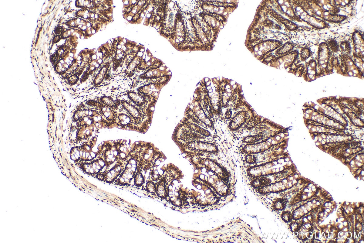
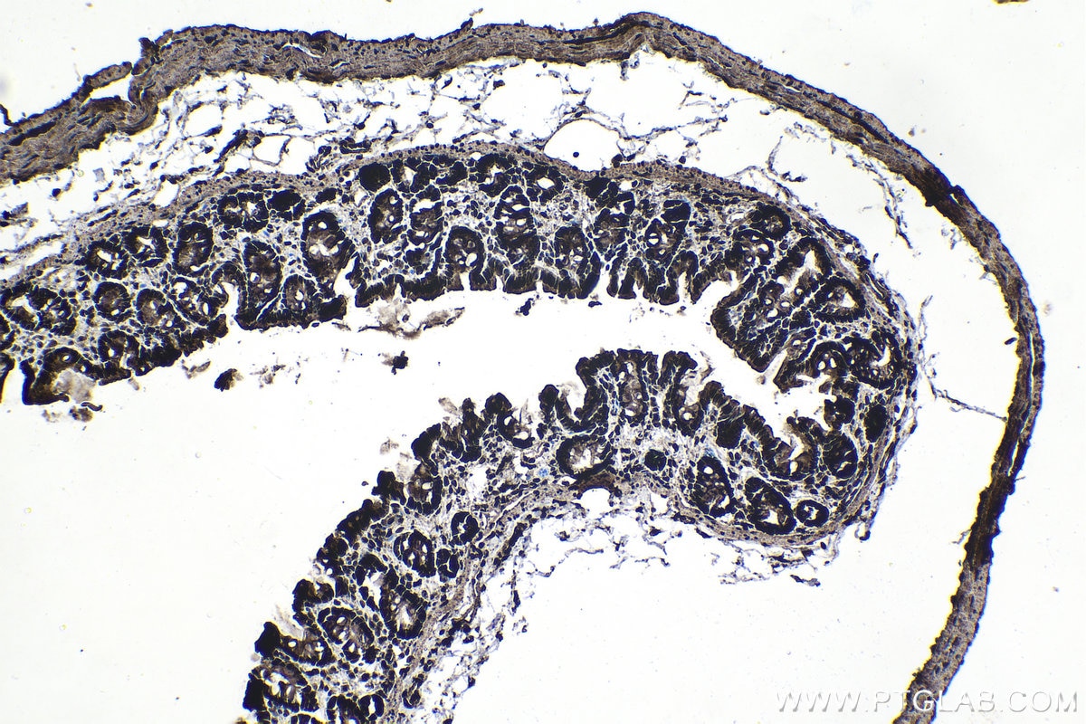
IHC staining of mouse colon using 25232-1-AP (same clone as 25232-1-PBS)
Immunohistochemical analysis of paraffin-embedded mouse colon tissue slide using 25232-1-AP (CCAR1 antibody) at dilution of 1:200 (under 10x lens). Heat mediated antigen retrieval with Tris-EDTA buffer (pH 9.0). This data was developed using the same antibody clone with 25232-1-PBS in a different storage buffer formulation.
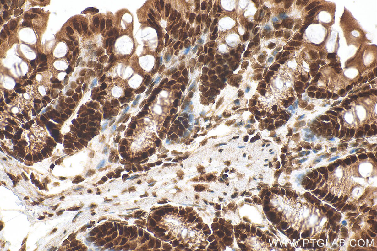
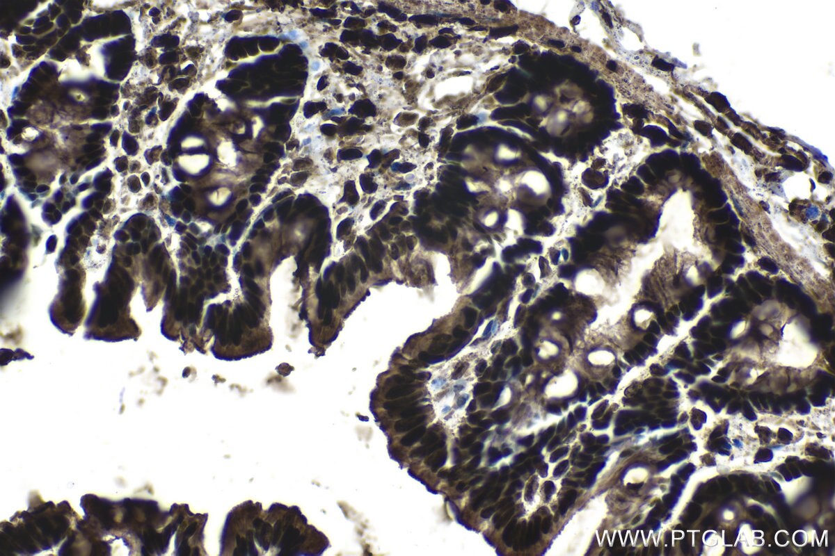
IHC staining of mouse colon using 25232-1-AP (same clone as 25232-1-PBS)
Immunohistochemical analysis of paraffin-embedded mouse colon tissue slide using 25232-1-AP (CCAR1 antibody) at dilution of 1:200 (under 40x lens). Heat mediated antigen retrieval with Tris-EDTA buffer (pH 9.0). This data was developed using the same antibody clone with 25232-1-PBS in a different storage buffer formulation.
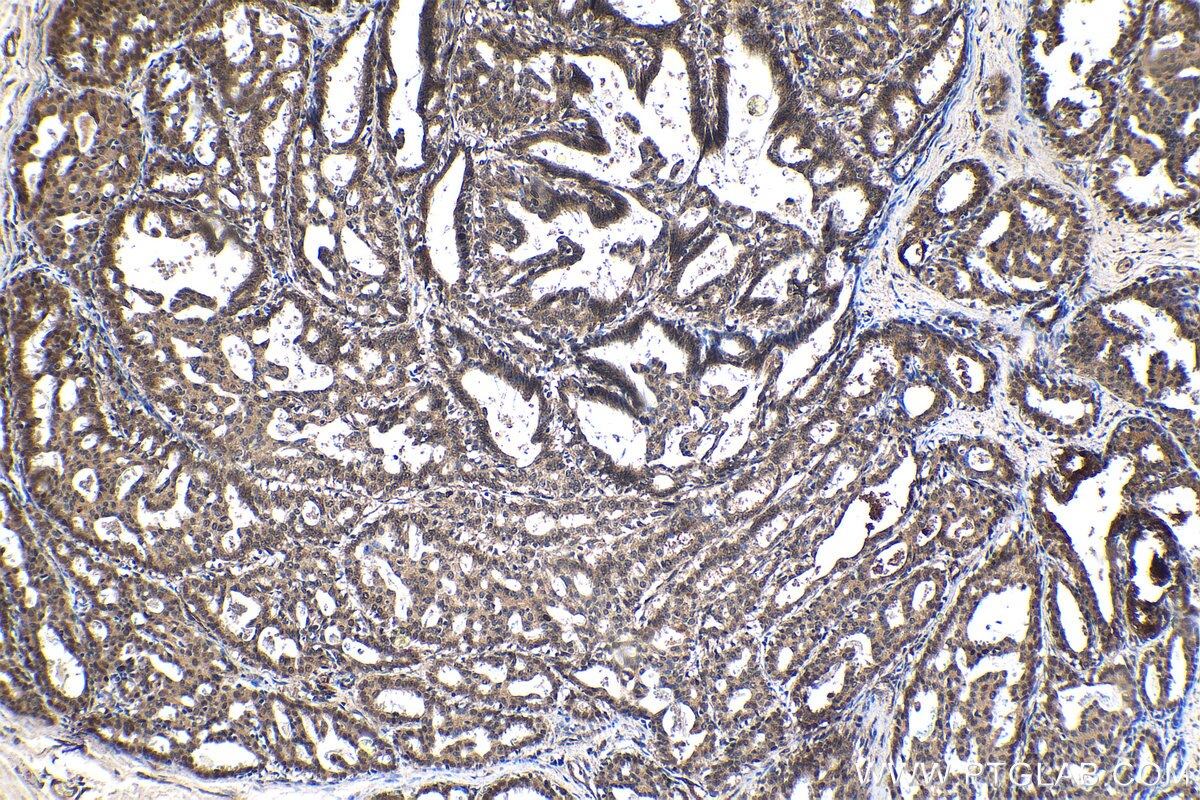
IHC staining of human breast cancer using 25232-1-AP (same clone as 25232-1-PBS)
Immunohistochemical analysis of paraffin-embedded human breast cancer tissue slide using 25232-1-AP (CCAR1 antibody) at dilution of 1:200 (under 10x lens). This data was developed using the same antibody clone with 25232-1-PBS in a different storage buffer formulation.
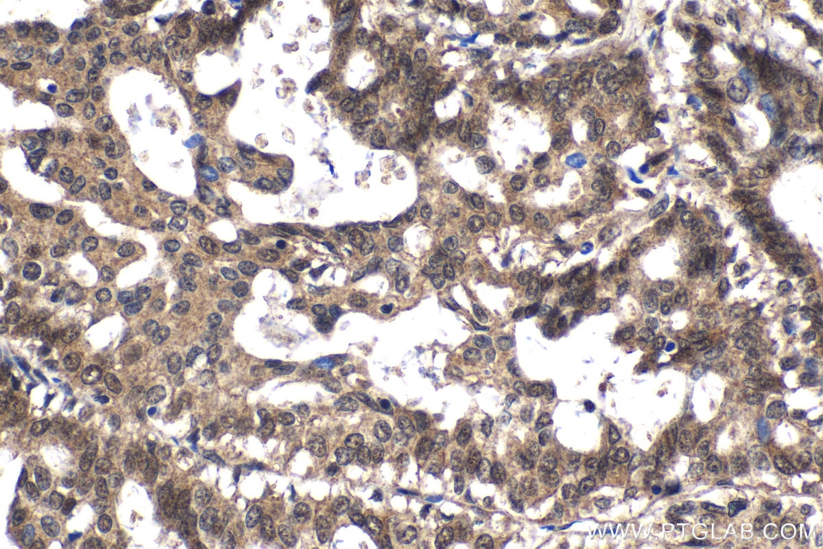
IHC staining of human breast cancer using 25232-1-AP (same clone as 25232-1-PBS)
Immunohistochemical analysis of paraffin-embedded human breast cancer tissue slide using 25232-1-AP (CCAR1 antibody) at dilution of 1:200 (under 40x lens). This data was developed using the same antibody clone with 25232-1-PBS in a different storage buffer formulation.
IHC staining of rat colon using 25232-1-AP (same clone as 25232-1-PBS)
Immunohistochemical analysis of paraffin-embedded rat colon tissue slide using 25232-1-AP (CCAR1 antibody) at dilution of 1:200 (under 10x lens). Heat mediated antigen retrieval with Tris-EDTA buffer (pH 9.0). This data was developed using the same antibody clone with 25232-1-PBS in a different storage buffer formulation.
IHC staining of rat colon using 25232-1-AP (same clone as 25232-1-PBS)
Immunohistochemical analysis of paraffin-embedded rat colon tissue slide using 25232-1-AP (CCAR1 antibody) at dilution of 1:200 (under 40x lens). Heat mediated antigen retrieval with Tris-EDTA buffer (pH 9.0). This data was developed using the same antibody clone with 25232-1-PBS in a different storage buffer formulation.
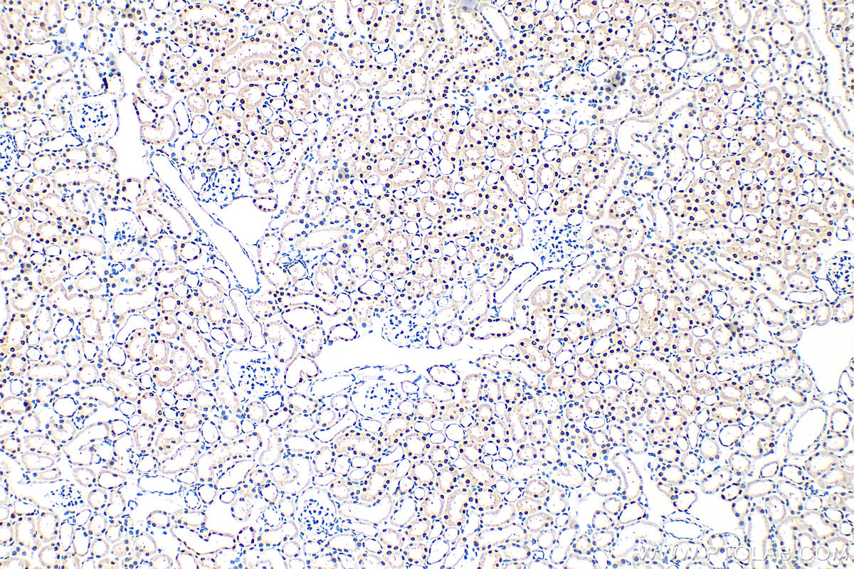
IHC staining of rat kidney using 25232-1-AP (same clone as 25232-1-PBS)
Immunohistochemical analysis of paraffin-embedded rat kidney tissue slide using 25232-1-AP (CCAR1 antibody) at dilution of 1:200 (under 10x lens). Heat mediated antigen retrieval with Tris-EDTA buffer (pH 9.0). This data was developed using the same antibody clone with 25232-1-PBS in a different storage buffer formulation.
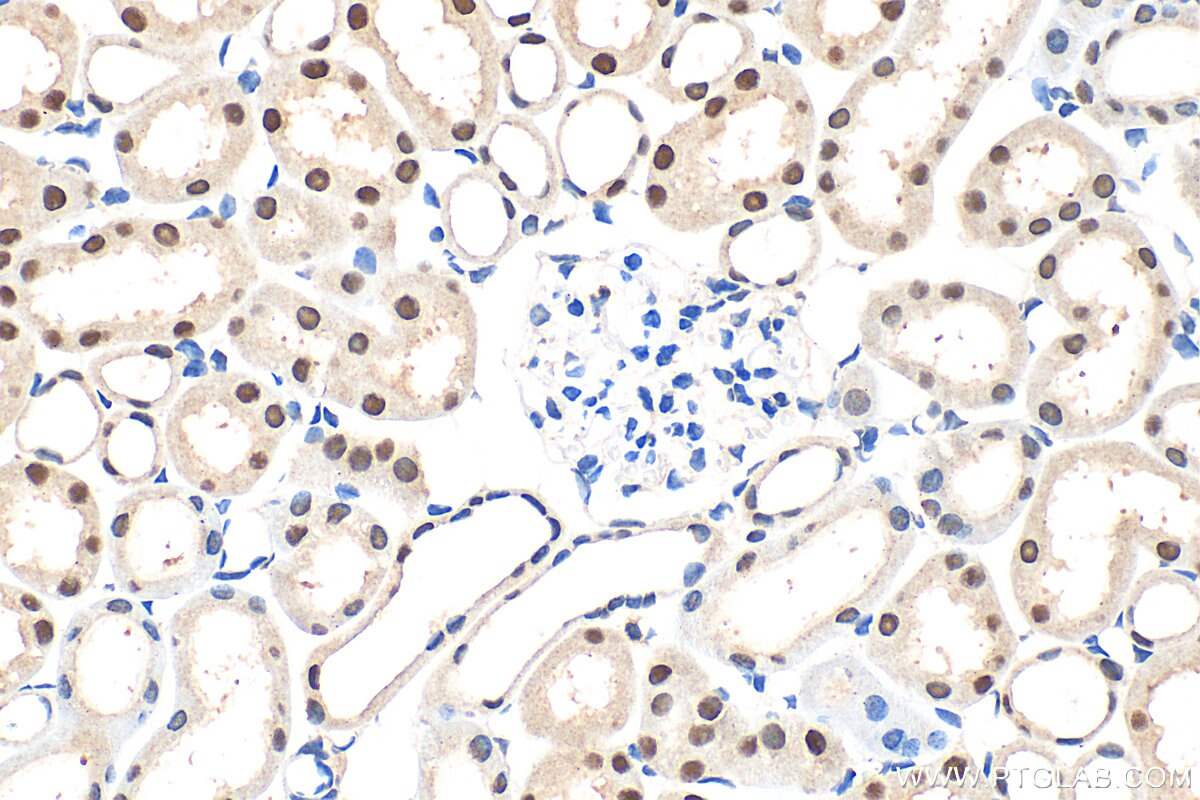
IHC staining of rat kidney using 25232-1-AP (same clone as 25232-1-PBS)
Immunohistochemical analysis of paraffin-embedded rat kidney tissue slide using 25232-1-AP (CCAR1 antibody) at dilution of 1:200 (under 40x lens). Heat mediated antigen retrieval with Tris-EDTA buffer (pH 9.0). This data was developed using the same antibody clone with 25232-1-PBS in a different storage buffer formulation.
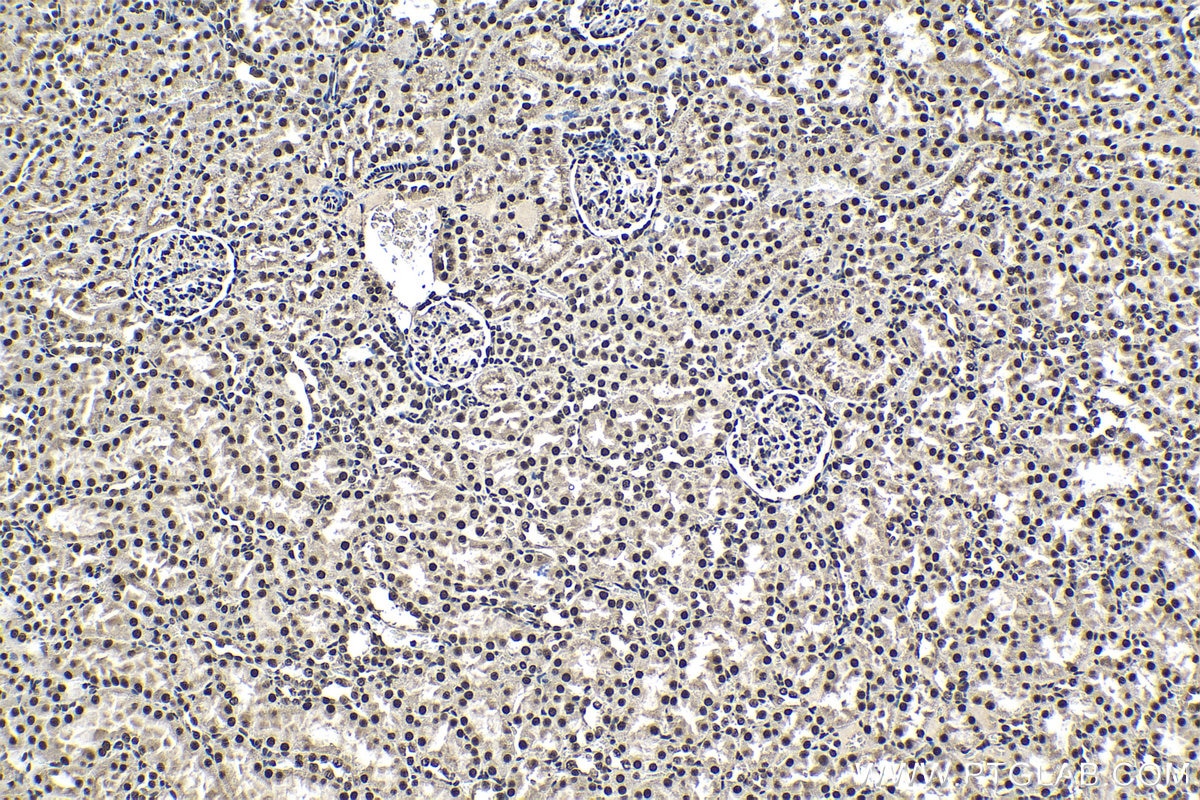
IHC staining of mouse kidney using 25232-1-AP (same clone as 25232-1-PBS)
Immunohistochemical analysis of paraffin-embedded mouse kidney tissue slide using 25232-1-AP (CCAR1 antibody) at dilution of 1:200 (under 10x lens). Heat mediated antigen retrieval with Tris-EDTA buffer (pH 9.0). This data was developed using the same antibody clone with 25232-1-PBS in a different storage buffer formulation.
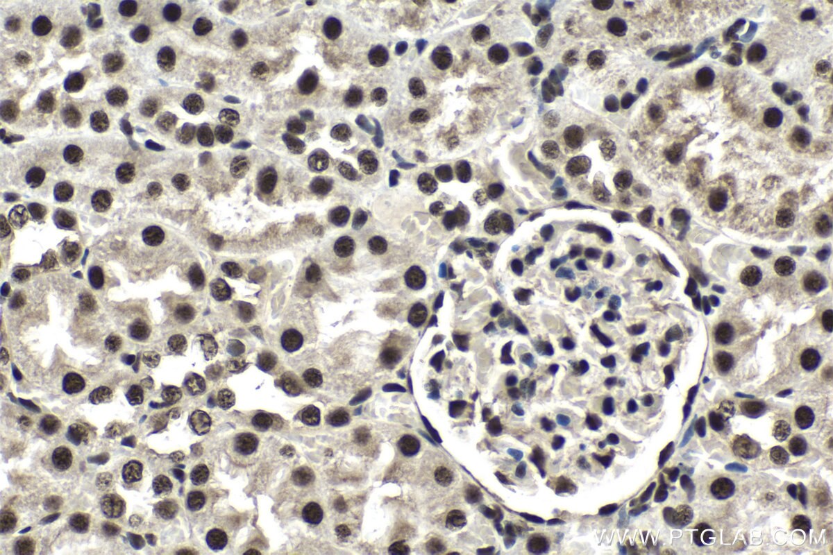
IHC staining of mouse kidney using 25232-1-AP (same clone as 25232-1-PBS)
Immunohistochemical analysis of paraffin-embedded mouse kidney tissue slide using 25232-1-AP (CCAR1 antibody) at dilution of 1:200 (under 40x lens). Heat mediated antigen retrieval with Tris-EDTA buffer (pH 9.0). This data was developed using the same antibody clone with 25232-1-PBS in a different storage buffer formulation.
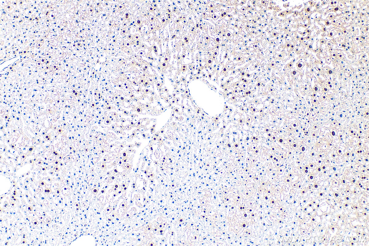
IHC staining of mouse liver using 25232-1-AP (same clone as 25232-1-PBS)
Immunohistochemical analysis of paraffin-embedded mouse liver tissue slide using 25232-1-AP (CCAR1 antibody) at dilution of 1:200 (under 10x lens). Heat mediated antigen retrieval with Tris-EDTA buffer (pH 9.0). This data was developed using the same antibody clone with 25232-1-PBS in a different storage buffer formulation.
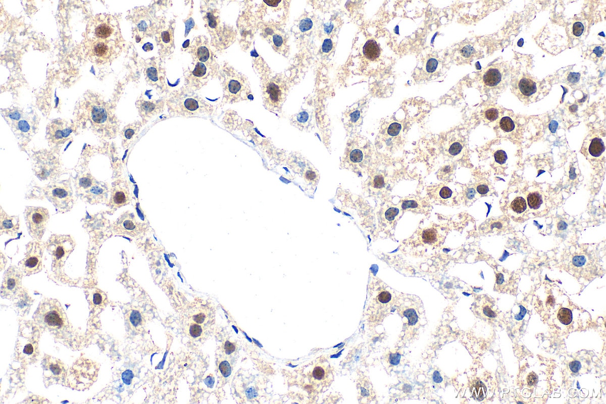
IHC staining of mouse liver using 25232-1-AP (same clone as 25232-1-PBS)
Immunohistochemical analysis of paraffin-embedded mouse liver tissue slide using 25232-1-AP (CCAR1 antibody) at dilution of 1:200 (under 40x lens). Heat mediated antigen retrieval with Tris-EDTA buffer (pH 9.0). This data was developed using the same antibody clone with 25232-1-PBS in a different storage buffer formulation.
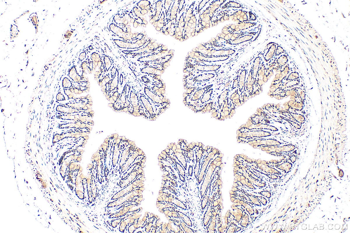
IHC staining of mouse colon using 25232-1-AP (same clone as 25232-1-PBS)
Immunohistochemical analysis of paraffin-embedded mouse colon tissue slide using 25232-1-AP (CCAR1 antibody) at dilution of 1:200 (under 10x lens). Heat mediated antigen retrieval with Tris-EDTA buffer (pH 9.0). This data was developed using the same antibody clone with 25232-1-PBS in a different storage buffer formulation.
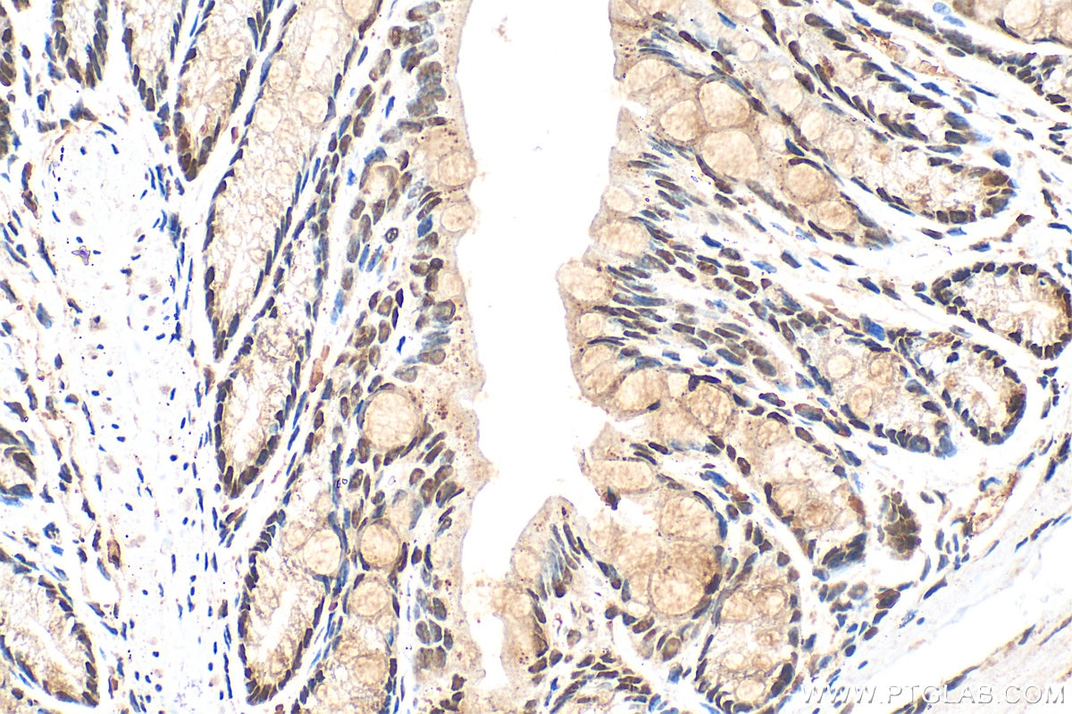
IHC staining of mouse colon using 25232-1-AP (same clone as 25232-1-PBS)
Immunohistochemical analysis of paraffin-embedded mouse colon tissue slide using 25232-1-AP (CCAR1 antibody) at dilution of 1:200 (under 40x lens). Heat mediated antigen retrieval with Tris-EDTA buffer (pH 9.0). This data was developed using the same antibody clone with 25232-1-PBS in a different storage buffer formulation.
IHC staining of human colon using 25232-1-AP (same clone as 25232-1-PBS)
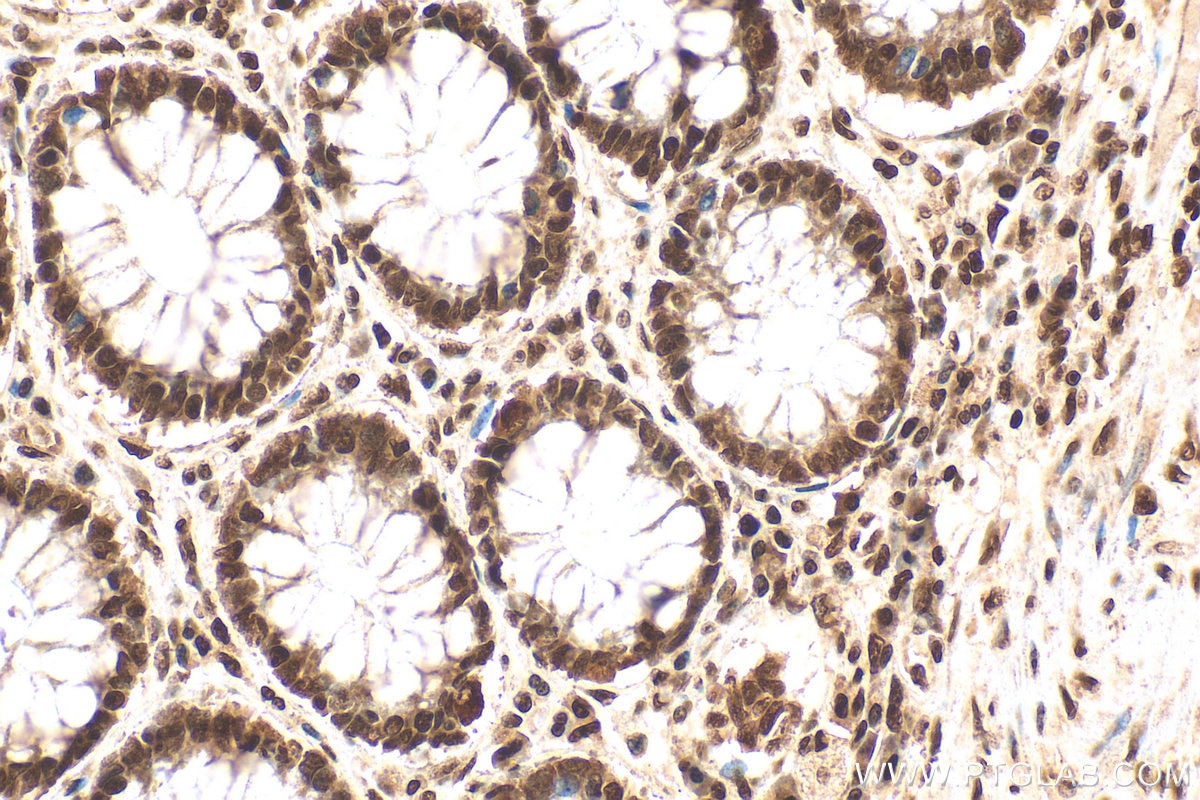
Immunohistochemical analysis of paraffin-embedded human colon tissue slide using 25232-1-AP (CCAR1 antibody) at dilution of 1:200 (under 40x lens). Heat mediated antigen retrieval with Tris-EDTA buffer (pH 9.0). This data was developed using the same antibody clone with 25232-1-PBS in a different storage buffer formulation.
IHC staining of human colon using 25232-1-AP (same clone as 25232-1-PBS)
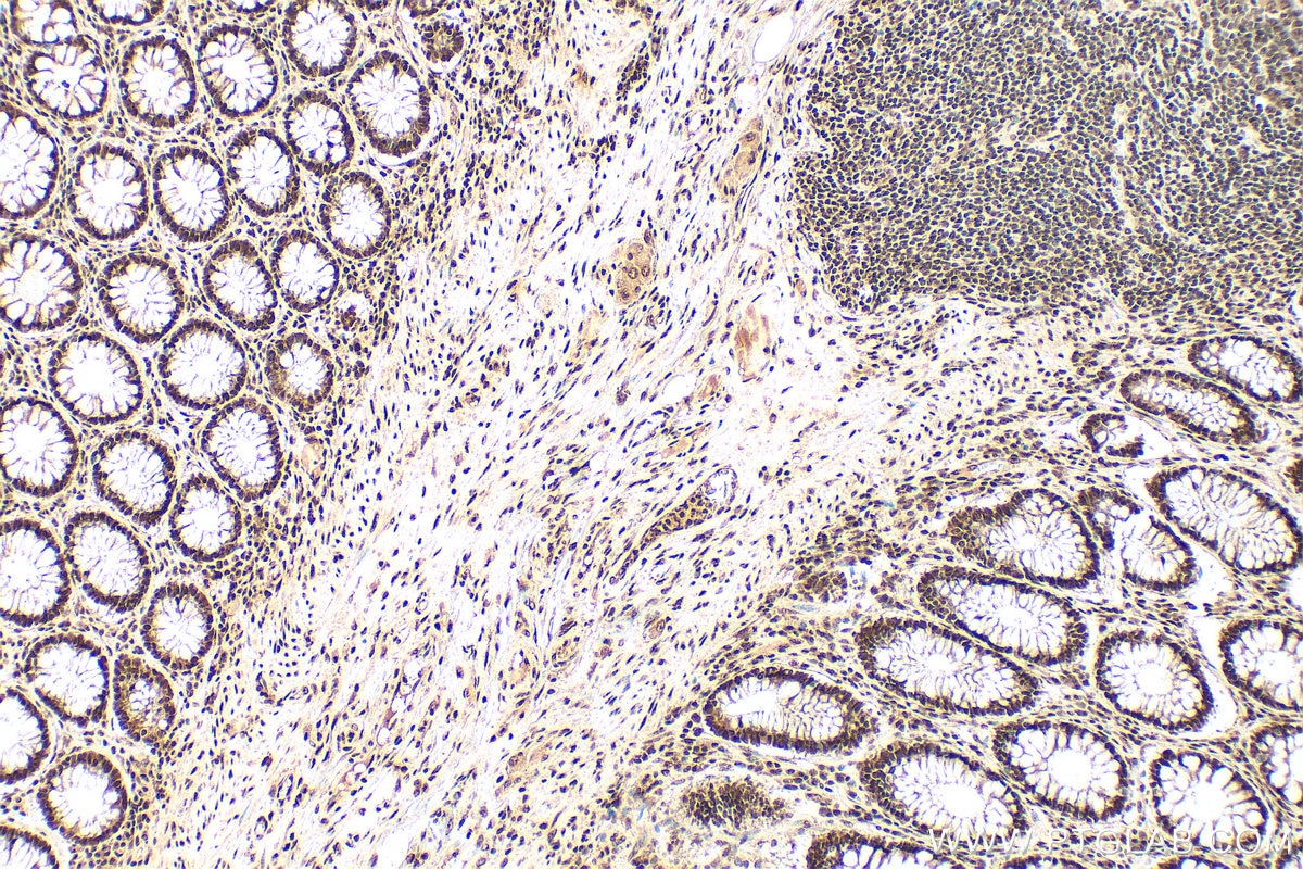
Immunohistochemical analysis of paraffin-embedded human colon tissue slide using 25232-1-AP (CCAR1 antibody) at dilution of 1:200 (under 10x lens). Heat mediated antigen retrieval with Tris-EDTA buffer (pH 9.0). This data was developed using the same antibody clone with 25232-1-PBS in a different storage buffer formulation.
IF/ICC Figures
IF Staining of A431 using 25232-1-AP (same clone as 25232-1-PBS)
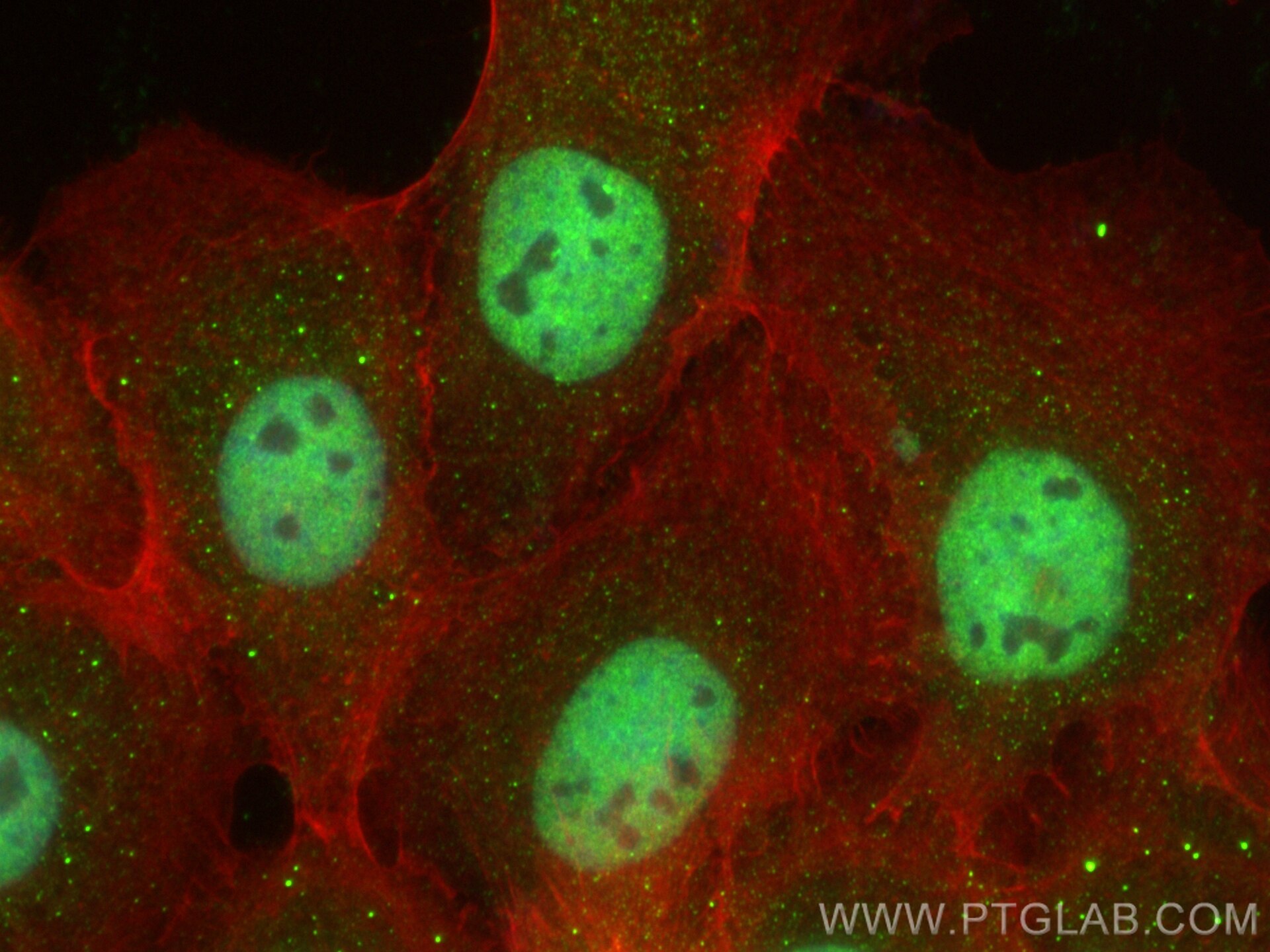
Immunofluorescent analysis of (4% PFA) fixed A431 cells using CCAR1 antibody (25232-1-AP) at dilution of 1:200 and Multi-rAb CoraLite ® Plus 488-Goat Anti-Rabbit Recombinant Secondary Antibody (H+L) (RGAR002), CL594-phalloidin (red). This data was developed using the same antibody clone with 25232-1-PBS in a different storage buffer formulation.
IF Staining of A431 using 25232-1-AP (same clone as 25232-1-PBS)
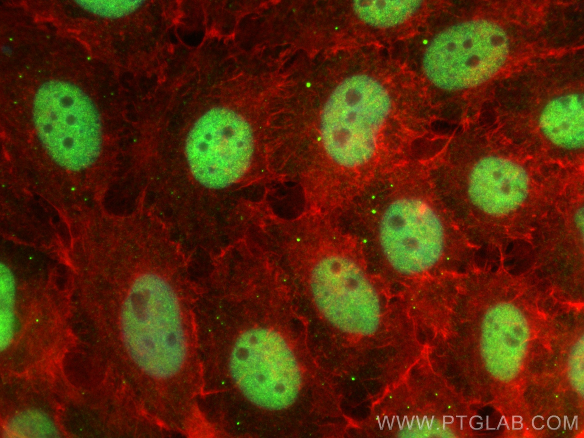
Immunofluorescent analysis of (4% PFA) fixed A431 cells using CCAR1 antibody (25232-1-AP) at dilution of 1:200 and Multi-rAb CoraLite ® Plus 488-Goat Anti-Rabbit Recombinant Secondary Antibody (H+L) (RGAR002), CL594-phalloidin (red). This data was developed using the same antibody clone with 25232-1-PBS in a different storage buffer formulation.