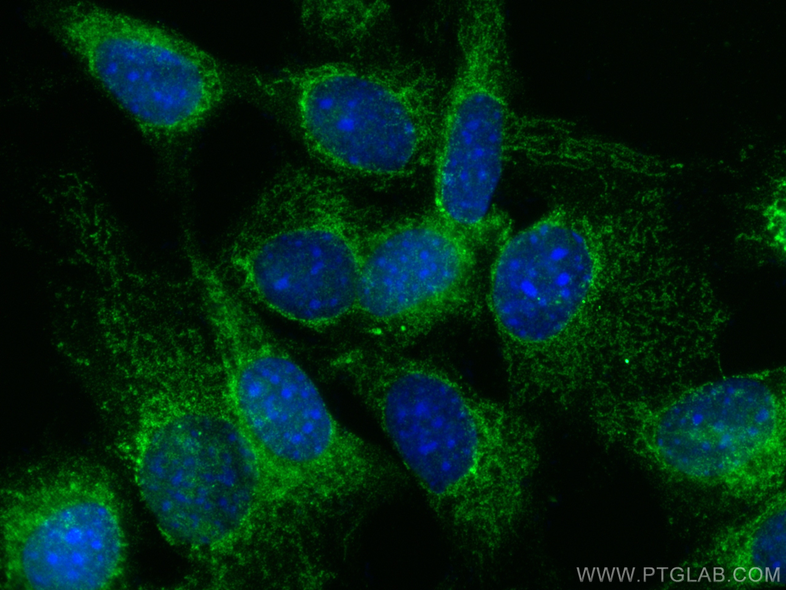
Validation Data Gallery
Tested Applications
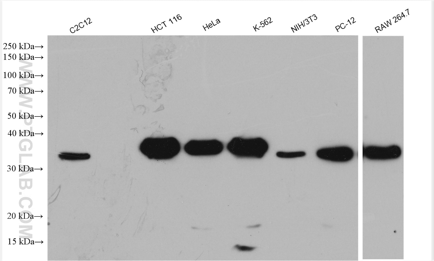
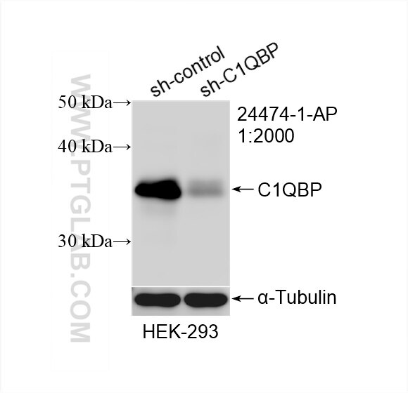
| Positive WB detected in | C2C12 cells, HEK-293 cells, HCT 116 cells, HeLa cells, K-562 cells, NIH/3T3 cells, PC-12 cells, RAW 264.7 cells |
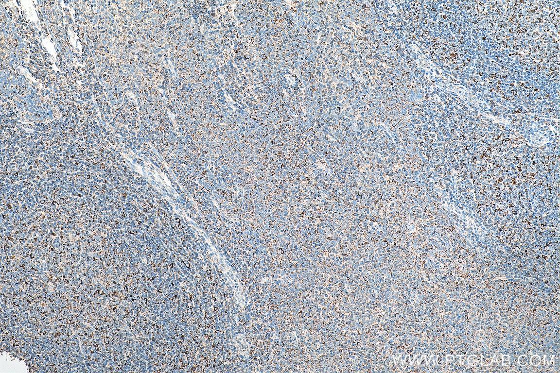
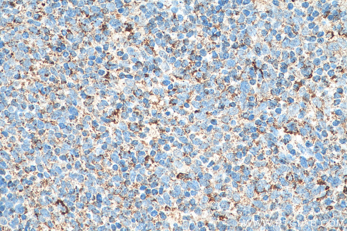
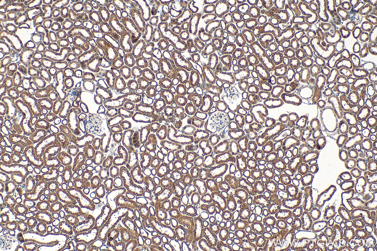
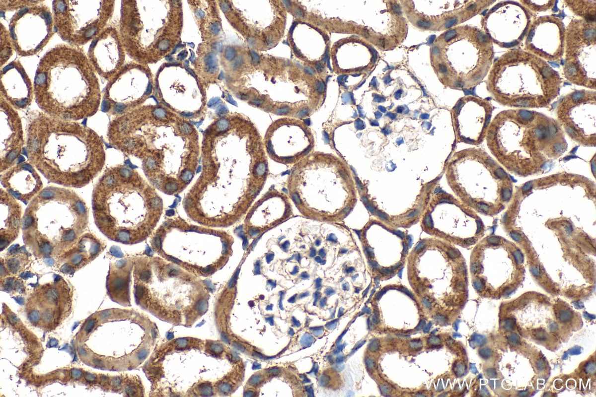
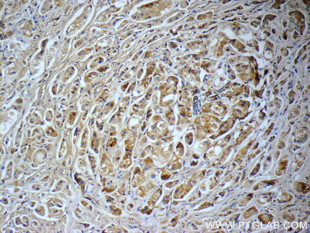
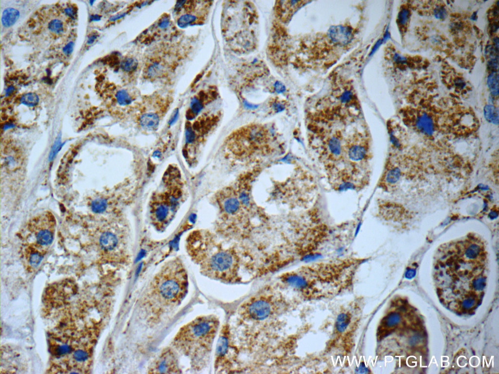
| Positive IHC detected in | human tonsillitis tissue, human breast cancer tissue, mouse kidney tissue Note: suggested antigen retrieval with TE buffer pH 9.0; (*) Alternatively, antigen retrieval may be performed with citrate buffer pH 6.0 |
| Positive IF/ICC detected in | NIH/3T3 cells, HeLa cells |
Recommended dilution
| Application | Dilution |
|---|---|
| Western Blot (WB) | WB : 1:5000-1:50000 |
| Immunohistochemistry (IHC) | IHC : 1:50-1:500 |
| Immunofluorescence (IF)/ICC | IF/ICC : 1:50-1:500 |
| It is recommended that this reagent should be titrated in each testing system to obtain optimal results. | |
| Sample-dependent, Check data in validation data gallery. | |
Published Applications
| KD/KO | See 3 publications below |
| WB | See 6 publications below |
| IHC | See 2 publications below |
| IF | See 5 publications below |
Product Information
24474-1-AP targets C1QBP in WB, IHC, IF/ICC, ELISA applications and shows reactivity with human, mouse, rat samples.
| Tested Reactivity | human, mouse, rat |
| Cited Reactivity | human, mouse, rat |
| Host / Isotype | Rabbit / IgG |
| Class | Polyclonal |
| Type | Antibody |
| Immunogen |
CatNo: Ag19773 Product name: Recombinant human C1QBP protein Source: e coli.-derived, PGEX-4T Tag: GST Domain: 1-282 aa of BC013731 Sequence: MLPLLRCVPRVLGSSVAGLRAAAPASPFRQLLQPAPRLCTRPFGLLSVRAGSERRPGLLRPRGPCACGCGCGSLHTDGDKAFVDFLSDEIKEERKIQKHKTLPKMSGGWELELNGTEAKLVRKVAGEKITVTFNINNSIPPTFDGEEEPSQGQKVEEQEPELTSTPNFVVEVIKNDDGKKALVLDCHYPEDEVGQEDEAESDIFSIREVSFQSTGESEWKDTNYTLNTDSLDWALYDHLMDFLADRGVDNTFADELVELSTALEHQEYITFLEDLKSFVKSQ 相同性解析による交差性が予測される生物種 |
| Full Name | complement component 1, q subcomponent binding protein |
| Calculated molecular weight | 282 aa, 31 kDa |
| Observed molecular weight | 32-35 kDa |
| GenBank accession number | BC013731 |
| Gene Symbol | C1QBP |
| Gene ID (NCBI) | 708 |
| RRID | AB_2827427 |
| Conjugate | Unconjugated |
| Form | |
| Form | Liquid |
| Purification Method | Antigen affinity purification |
| UNIPROT ID | Q07021 |
| Storage Buffer | PBS with 0.02% sodium azide and 50% glycerol{{ptg:BufferTemp}}7.3 |
| Storage Conditions | Store at -20°C. Stable for one year after shipment. Aliquoting is unnecessary for -20oC storage. |
Background Information
C1QBP, also named as gC1q receptor (gC1qR), p32, p33, and hyaluronan-binding protein 1 (HABP1), is a protein initially copurified with splicing factor SF2 (PMID: 1830244). The protein is synthesized as a pro-protein of 282 amino acids (aa) that is post-translationally processed by removal of the initial 73 aa to a mature protein of 209 aa (PMID: 8262387). C1QBP is an evolutionary conserved and ubiquitously expressed multifunctional protein and has been reported to be a predominantly mitochondrial matrix protein involved in inflammation and infection processes, mitochondrial ribosome biogenesis, regulation of apoptosis and nuclear transcription, and pre-mRNA splicing (PMID: 28942965).
Protocols
| Product Specific Protocols | |
|---|---|
| IF protocol for C1QBP antibody 24474-1-AP | Download protocol |
| IHC protocol for C1QBP antibody 24474-1-AP | Download protocol |
| WB protocol for C1QBP antibody 24474-1-AP | Download protocol |
| Standard Protocols | |
|---|---|
| Click here to view our Standard Protocols |
Publications
| Species | Application | Title |
|---|---|---|
Blood C1Q labels a highly aggressive macrophage-like leukemia population indicating extramedullary infiltration and relapse
| ||
Proc Natl Acad Sci U S A A nucleolar long "non-coding" RNA encodes a novel protein that functions in response to stress | ||
J Control Release PSMA-targeted nanoparticles for specific penetration of blood-brain tumor barrier and combined therapy of brain metastases. | ||
J Ethnopharmacol Exploring the therapeutic effects and molecular mechanisms of total flavonoids of Abelmoschus manihot (L.) Medic in the treatment of IgA nephropathy based on WGCNA | ||
Cancer Sci Complement C1q binding protein regulates T cells' mitochondrial fitness to affect their survival, proliferation, and anti-tumor immune function.
| ||
Int J Mol Sci The Expression Pattern of p32 in Sheep Muscle and Its Role in Differentiation, Cell Proliferation, and Apoptosis of Myoblasts.
|

