PELP1 Monoclonal antibody
PELP1 Monoclonal Antibody for WB, IF, IHC, ELISA, FC (Intra)
Host / Isotype
Mouse / IgG2b
Reactivity
Human and More (1)
Applications
WB, IF, IHC, ELISA, FC (Intra)
Conjugate
Unconjugated
CloneNo.
1B11E4
Cat no : 67050-1-Ig
Synonyms
Validation Data Gallery
 at dilution of 1:5000 incubated at room temperature for 1.5 hours.")
 at dilution of 1:5000 incubated at room temperature for 1.5 hours.")
 at dilution of 1:5000 incubated at room temperature for 1.5 hours.")
 at dilution of 1:1000 (under 10x lens. Heat mediated antigen retrieval with Tris-EDTA buffer (pH 9.0).")
 at dilution of 1:1000 (under 40x lens. Heat mediated antigen retrieval with Tris-EDTA buffer (pH 9.0).")
 at dilution of 1:600 (under 10x lens. Heat mediated antigen retrieval with Tris-EDTA buffer (pH 9.0).")
 at dilution of 1:600 (under 40x lens. Heat mediated antigen retrieval with Tris-EDTA buffer (pH 9.0).")
 fixed human breast cancer tissue using <a class='green' href='/productredirect?CatalogNo=PELP1' target='_blank'>PELP1</a> antibody (<a class='green' href='/productredirect?CatalogNo=67050-1-Ig' target='_blank'>67050-1-Ig</a>, Clone: 1<a class='green' href='/productredirect?CatalogNo=B11' target='_blank'>B11</a><a class='green' href='/productredirect?CatalogNo=E4' target='_blank'>E4</a> ) at dilution of 1:400 and CoraLite®488-Conjugated AffiniPure Goat Anti-Mouse IgG(H+L).")
 fixed human breast cancer tissue using <a class='green' href='/productredirect?CatalogNo=PELP1' target='_blank'>PELP1</a> antibody (<a class='green' href='/productredirect?CatalogNo=67050-1-Ig' target='_blank'>67050-1-Ig</a>, Clone: 1<a class='green' href='/productredirect?CatalogNo=B11' target='_blank'>B11</a><a class='green' href='/productredirect?CatalogNo=E4' target='_blank'>E4</a> ) at dilution of 1:400 and CoraLite®488-Conjugated AffiniPure Goat Anti-Mouse IgG(H+L).")
 and CoraLite®488-Conjugated AffiniPure Goat Anti-Mouse IgG(H+L) at dilution 1:1000 (red), or 0.4 ug Mouse <a class='green' href='/productredirect?CatalogNo=IgG2' target='_blank'>IgG2</a>b Isotype Control (<a class='green' href='/productredirect?CatalogNo=66360-3-Ig' target='_blank'>66360-3-Ig</a>, Clone: <a class='green' href='/productredirect?CatalogNo=K11' target='_blank'>K11</a><a class='green' href='/productredirect?CatalogNo=B8' target='_blank'>B8</a><a class='green' href='/productredirect?CatalogNo=C4' target='_blank'>C4</a><a class='green' href='/productredirect?CatalogNo=B5' target='_blank'>B5</a>) (blue). Cells were fixed and permeabilized with Transcription Factor Staining Buffer Kit (<a class='green' href='/productredirect?CatalogNo=PF00011' target='_blank'>PF00011</a>).")
Tested Applications
| Positive WB detected in | HeLa cells, T-47D cells, Jurkat cells, HEK-293 cells, MCF-7 cells |
| Positive IHC detected in | human cervical cancer tissue, human breast cancer tissue Note: suggested antigen retrieval with TE buffer pH 9.0; (*) Alternatively, antigen retrieval may be performed with citrate buffer pH 6.0 |
| Positive IF detected in | human breast cancer tissue |
| Positive FC detected in | MCF-7 cells |
Recommended dilution
| Application | Dilution |
|---|---|
| Western Blot (WB) | WB : 1:2000-1:8000 |
| Immunohistochemistry (IHC) | IHC : 1:500-1:2000 |
| Immunofluorescence (IF) | IF : 1:200-1:800 |
| Flow Cytometry (FC) | FC : 0.40 ug per 10^6 cells in a 100 µl suspension |
| Sample-dependent, check data in validation data gallery | |
Published Applications
| WB | See 1 publications below |
| IHC | See 1 publications below |
Product Information
67050-1-Ig targets PELP1 in WB, IF, IHC, ELISA, FC (Intra) applications and shows reactivity with Human samples.
| Tested Reactivity | Human |
| Cited Reactivity | mouse |
| Host / Isotype | Mouse / IgG2b |
| Class | Monoclonal |
| Type | Antibody |
| Immunogen | PELP1 fusion protein Ag25729 相同性解析による交差性が予測される生物種 |
| Full Name | proline, glutamate and leucine rich protein 1 |
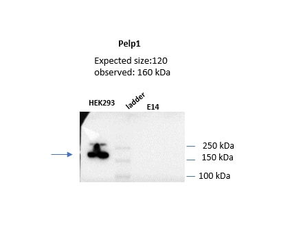
| Calculated molecular weight | 1130 aa, 120 kDa |
| Observed molecular weight | 160 kDa |
| GenBank accession number | BC069058 |
| Gene symbol | PELP1 |
| Gene ID (NCBI) | 27043 |
| RRID | AB_2882363 |
| Conjugate | Unconjugated |
| Form | Liquid |
| Purification Method | Protein A purification |
| Storage Buffer | PBS with 0.02% sodium azide and 50% glycerol pH 7.3. |
| Storage Conditions | Store at -20°C. Stable for one year after shipment. Aliquoting is unnecessary for -20oC storage. |
Background Information
PELP1 was first identified as a 160 kDa protein in a screen for Src homology 2 (SH2) domain-binding proteins. PELP1 is overexpressed in 60-80% of breast tumors and plays important roles in both ER genomic and non-genomic signaling. In vivo, PELP1 subcellular localization is primarily nuclear in normal breast tissue, but it is localized to the cytoplasm in about 40% of invasive breast tumors. In the nucleus, PELP1 interacts with a number of transcription factors. The proto-oncogenic functions of PELP1 involve different cellular processes including epigenetic modifications leading to ER transactivation and breast cancer progression. Furthermore, PELP1 activates kinase cascades in the cytoplasm such as MAPK activation via c-Src and PI3K signaling.
Protocols
| Product Specific Protocols | |
|---|---|
| WB protocol for PELP1 antibody 67050-1-Ig | Download protocol |
| IHC protocol for PELP1 antibody 67050-1-Ig | Download protocol |
| IF protocol for PELP1 antibody 67050-1-Ig | Download protocol |
| FC protocol for PELP1 antibody 67050-1-Ig | Download protocol |
| Standard Protocols | |
|---|---|
| Click here to view our Standard Protocols |
Publications
| Species | Application | Title |
|---|---|---|
Int Immunopharmacol MiR-195-5p is involved in testicular ischemia/reperfusion injury by directly targeting PELP1 and regulating spermatogonia pyroptosis |
Reviews
The reviews below have been submitted by verified Proteintech customers who received an incentive forproviding their feedback.
FH fatemeh (Verified Customer) (11-30-2021) | The antibody works well on human protein lystae (30 ug of protein from Hek293 cells, 8% gel, wet transfer 90min 300mA in tris-Gly buffer on PVDF, incubation with primary ab was onvernight at 4C). we coulnt see the specific band for mouse cells.
 |

