Midkine Polyclonal antibody
Midkine Polyclonal Antibody for WB, IHC, ELISA
Host / Isotype
Rabbit / IgG
Reactivity
Human, mouse
Applications
WB, IHC, ELISA
Conjugate
Unconjugated
Cat no : 28546-1-AP
Synonyms
Validation Data Gallery
Tested Applications
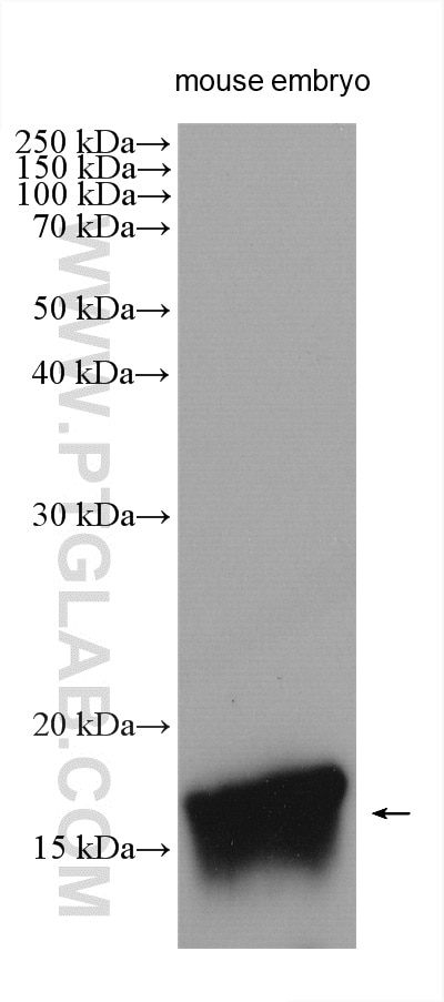
| Positive WB detected in | mouse embryo tissue |
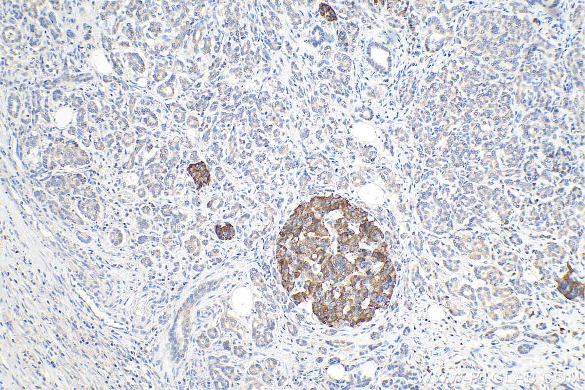
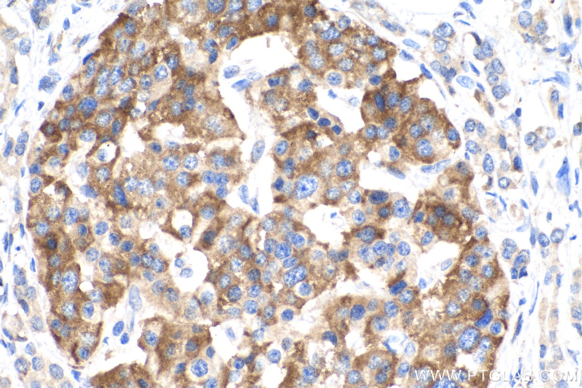
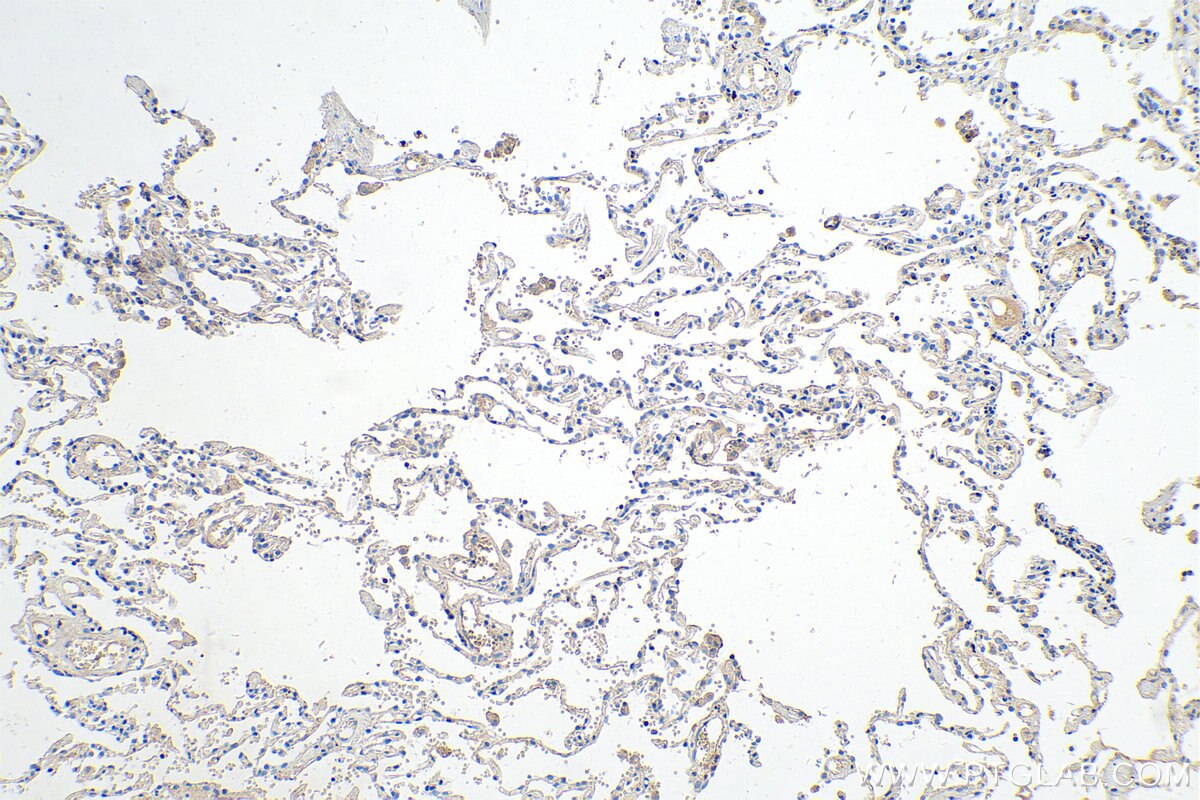
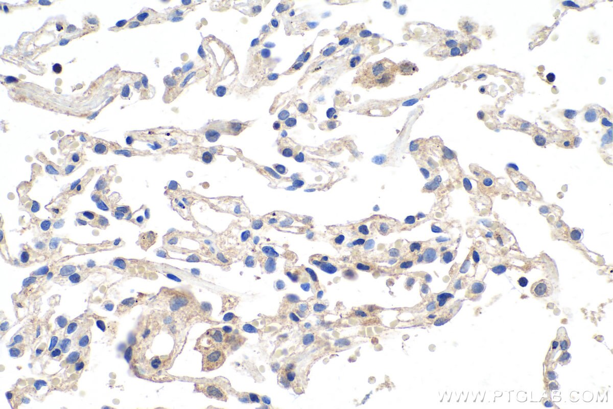
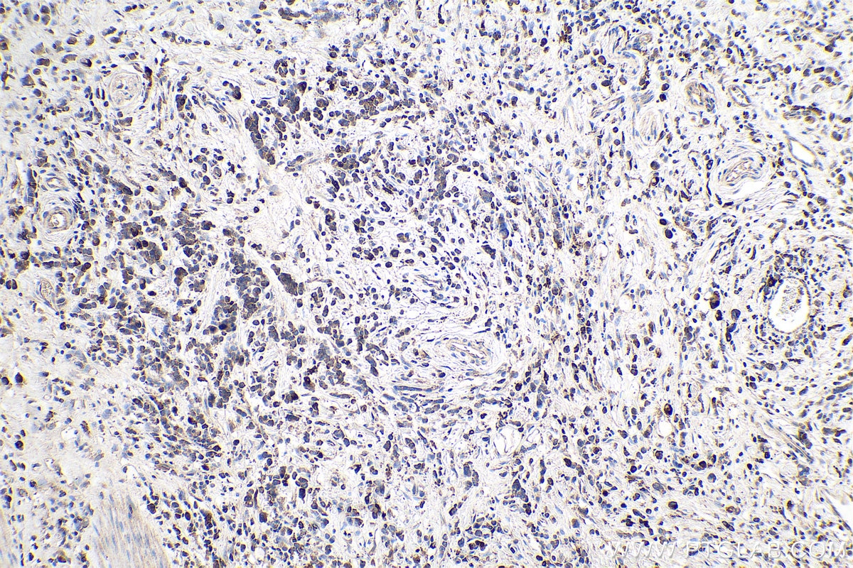
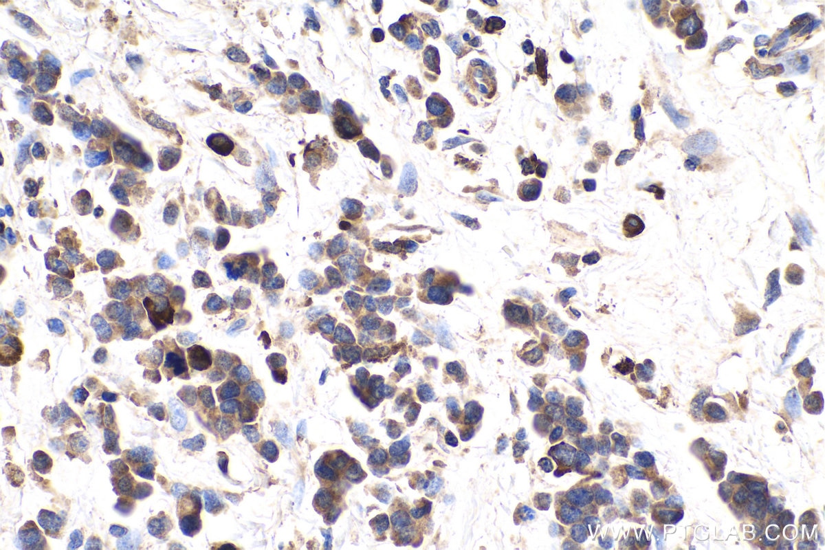
| Positive IHC detected in | human pancreas cancer tissue, human stomach cancer tissue, human lung cancer tissue Note: suggested antigen retrieval with TE buffer pH 9.0; (*) Alternatively, antigen retrieval may be performed with citrate buffer pH 6.0 |
Recommended dilution
| Application | Dilution |
|---|---|
| Western Blot (WB) | WB : 1:200-1:1000 |
| Immunohistochemistry (IHC) | IHC : 1:250-1:1000 |
| It is recommended that this reagent should be titrated in each testing system to obtain optimal results. | |
| Sample-dependent, Check data in validation data gallery. | |
Published Applications
| WB | See 1 publications below |
Product Information
28546-1-AP targets Midkine in WB, IHC, ELISA applications and shows reactivity with Human, mouse samples.
| Tested Reactivity | Human, mouse |
| Cited Reactivity | mouse |
| Host / Isotype | Rabbit / IgG |
| Class | Polyclonal |
| Type | Antibody |
| Immunogen | Midkine fusion protein Ag29185 相同性解析による交差性が予測される生物種 |
| Full Name | midkine (neurite growth-promoting factor 2) |
| Calculated molecular weight | 16 kDa |
| Observed molecular weight | 16 kDa |
| GenBank accession number | BC011704 |
| Gene symbol | Midkine |
| Gene ID (NCBI) | 4192 |
| RRID | AB_2881168 |
| Conjugate | Unconjugated |
| Form | Liquid |
| Purification Method | Antigen affinity purification |
| Storage Buffer | PBS with 0.02% sodium azide and 50% glycerol pH 7.3. |
| Storage Conditions | Store at -20°C. Stable for one year after shipment. Aliquoting is unnecessary for -20oC storage. |
Protocols
| Product Specific Protocols | |
|---|---|
| WB protocol for Midkine antibody 28546-1-AP | Download protocol |
| IHC protocol for Midkine antibody 28546-1-AP | Download protocol |
| Standard Protocols | |
|---|---|
| Click here to view our Standard Protocols |
Publications
| Species | Application | Title |
|---|---|---|
EBioMedicine Spatial and single-cell colocalisation analysis reveals MDK-mediated immunosuppressive environment with regulatory T cells in colorectal carcinogenesis |
