Validation Data Gallery
Tested Applications
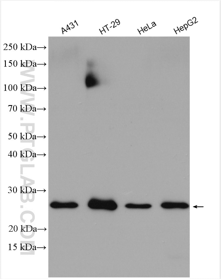
| Positive WB detected in | A431 cells, human preadipocyte cells, HT-29 cells, HeLa cells, HepG2 cells |
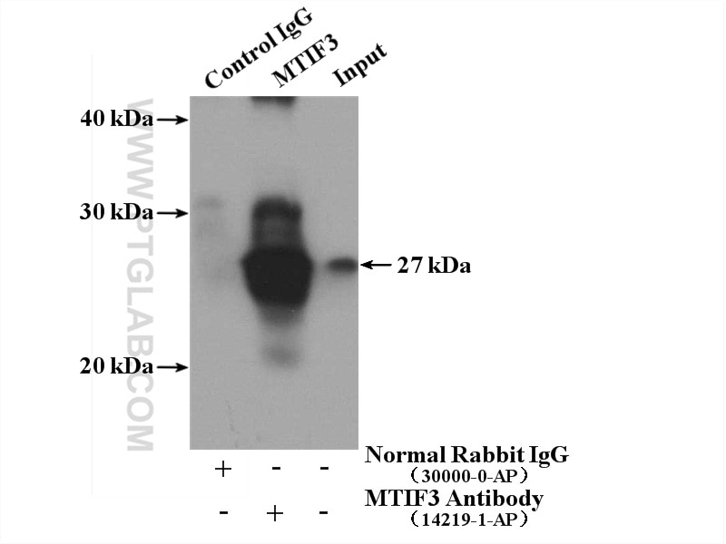
| Positive IP detected in | HeLa cells |
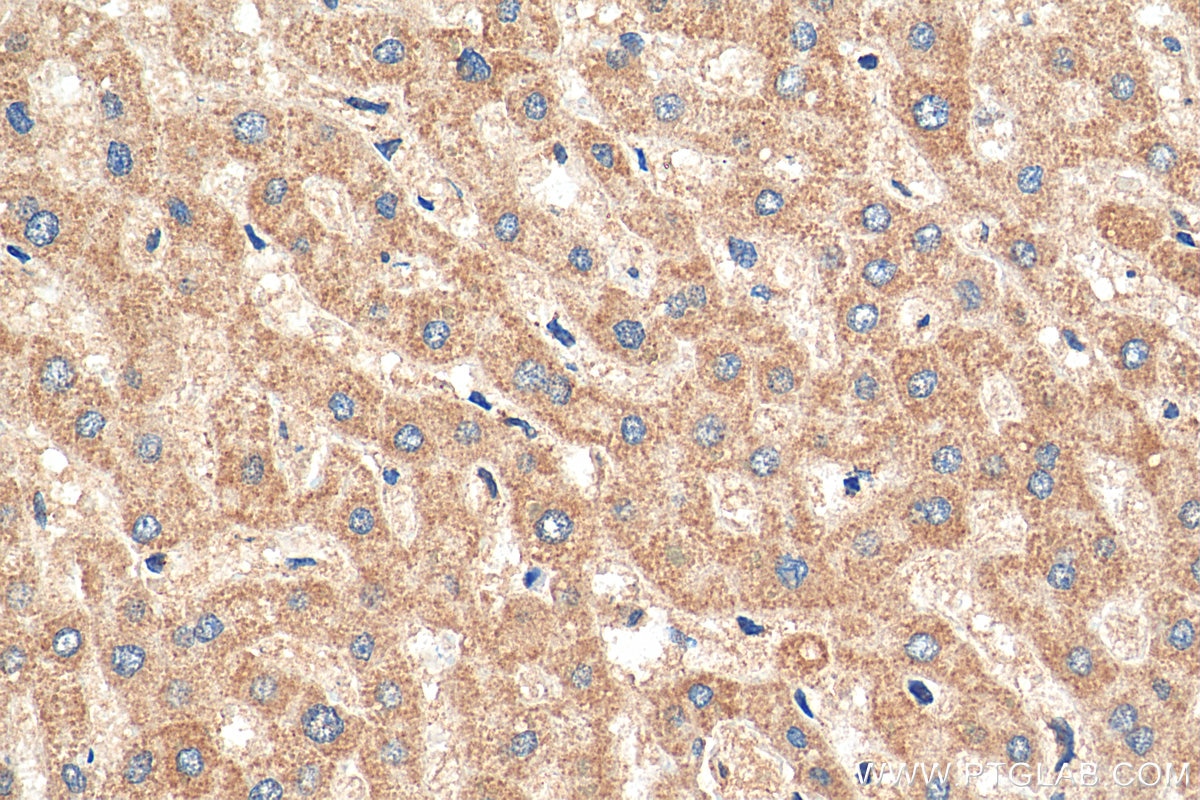
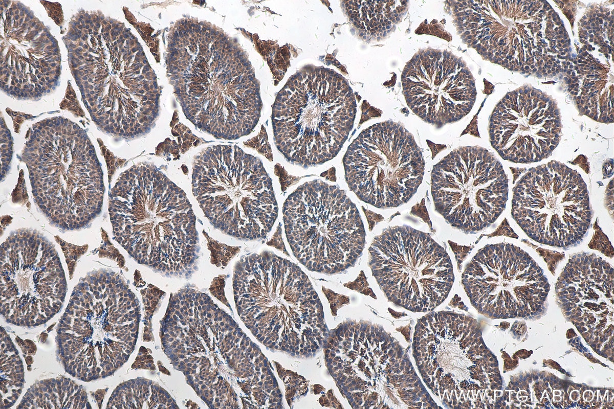
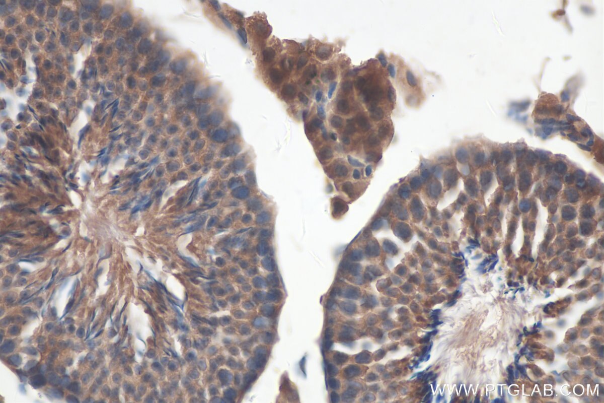
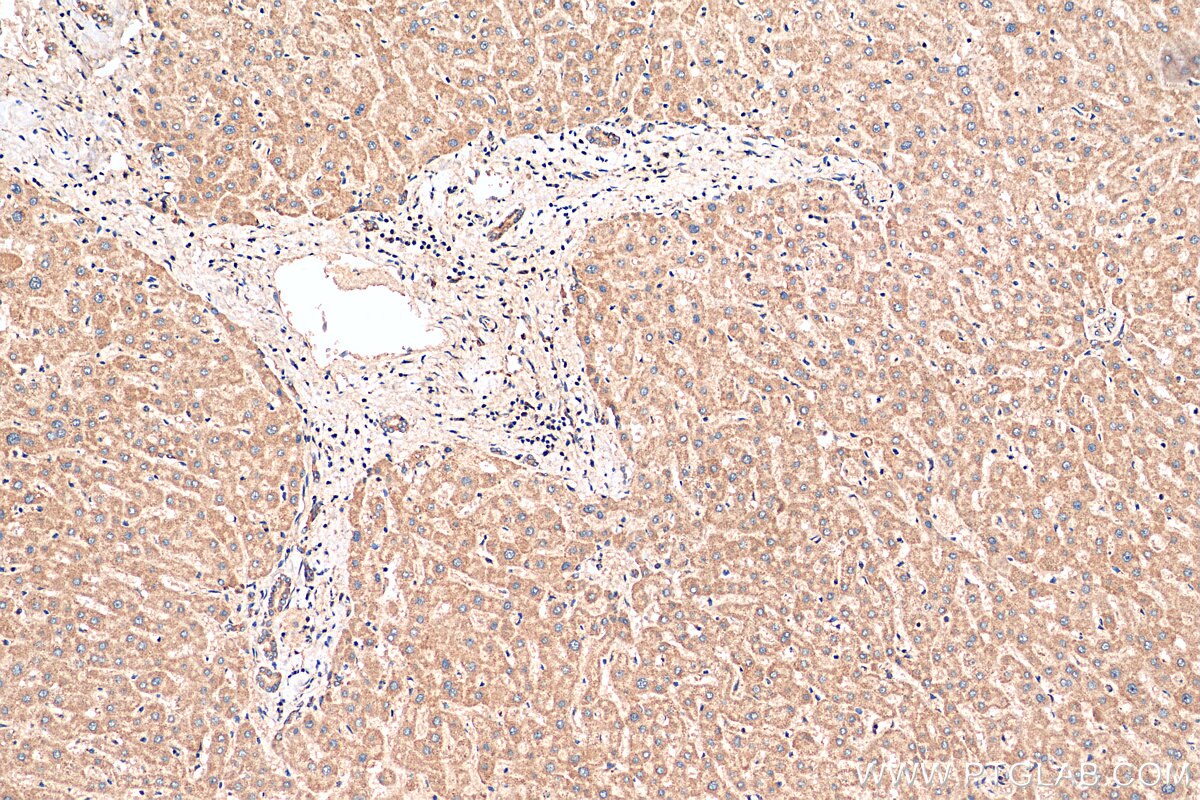
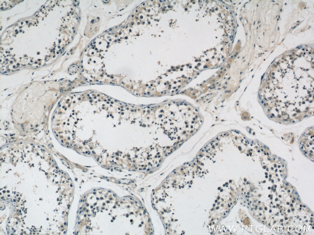
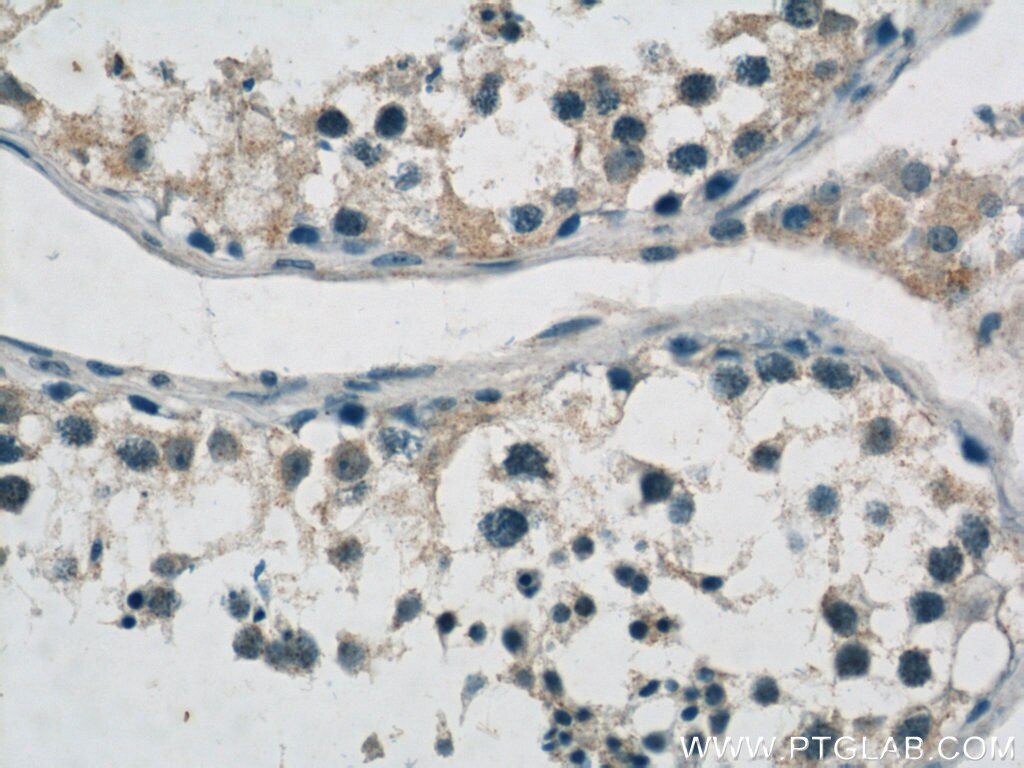
| Positive IHC detected in | human liver tissue, mouse testis tissue, human testis tissue Note: suggested antigen retrieval with TE buffer pH 9.0; (*) Alternatively, antigen retrieval may be performed with citrate buffer pH 6.0 |
Recommended dilution
| Application | Dilution |
|---|---|
| Western Blot (WB) | WB : 1:500-1:2000 |
| Immunoprecipitation (IP) | IP : 0.5-4.0 ug for 1.0-3.0 mg of total protein lysate |
| Immunohistochemistry (IHC) | IHC : 1:50-1:500 |
| It is recommended that this reagent should be titrated in each testing system to obtain optimal results. | |
| Sample-dependent, Check data in validation data gallery. | |
Published Applications
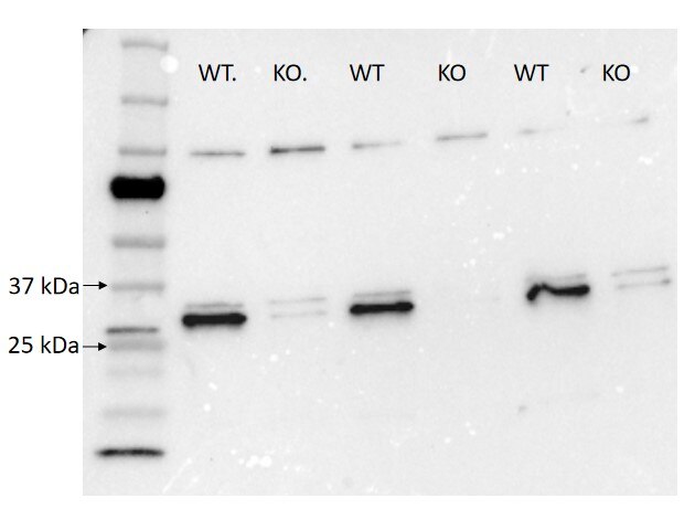
| WB | See 7 publications below |
Product Information
14219-1-AP targets MTIF3 in WB, IHC, IP, ELISA applications and shows reactivity with human, mouse samples.
| Tested Reactivity | human, mouse |
| Cited Reactivity | human, mouse, rat |
| Host / Isotype | Rabbit / IgG |
| Class | Polyclonal |
| Type | Antibody |
| Immunogen |
CatNo: Ag5457 Product name: Recombinant human MTIF3 protein Source: e coli.-derived, PGEX-4T Tag: GST Domain: 1-278 aa of BC046166 Sequence: MAALFLKRLTLQTVKSENSCIRCFGKHILQKTAPAQLSPIASAPRLSFLIHAKAFSTAEDTQNEGKKIKKNKTAFSNVGRKISQRVIHLFDEKGNDLGNMHRANVIRLMDERDLRLVQRNTSTEPAEYQLMTGLQILQERQRLREMEKANPKTGPTLRKELILSSNIGQHDLDTKTKQIQQWIKKKHLVQITIKKGKNVDVSENEMEEIFHQILQTMPGIATFSSRPQAVQGGKALMCVLRALSKNEEKAYKETQETQERDTLNKDHGNDKESNVLHQ 相同性解析による交差性が予測される生物種 |
| Full Name | mitochondrial translational initiation factor 3 |
| Calculated molecular weight | 32 kDa |
| Observed molecular weight | 29 kDa |
| GenBank accession number | BC046166 |
| Gene Symbol | MTIF3 |
| Gene ID (NCBI) | 219402 |
| RRID | AB_10638621 |
| Conjugate | Unconjugated |
| Form | |
| Form | Liquid |
| Purification Method | Antigen affinity purification |
| UNIPROT ID | Q9H2K0 |
| Storage Buffer | PBS with 0.02% sodium azide and 50% glycerol{{ptg:BufferTemp}}7.3 |
| Storage Conditions | Store at -20°C. Stable for one year after shipment. Aliquoting is unnecessary for -20oC storage. |
Background Information
MTIF3, also named as DC38, belongs to the IF-3 family. MTIF3 encodes a 29 kDa protein that promotes formation of the initiation complex on the mitochondrial 55S ribosome, thereby playing an active role in initiation of translation. Like bacterial IF3, MTIF3 is believed to bind first to the small mitoribosomal subunit to keep it dissociated from the large subunit during initiation. After binding of MTIF3, mRNA and formylated initiator methionyl-tRNA (fMet-tRNAifMet) bind to the small mitoribosomal subunit. The large subunit then joins the small subunit to form an elongation-competent ribosome (PMID:20887776, 31350787).
Protocols
| Product Specific Protocols | |
|---|---|
| IHC protocol for MTIF3 antibody 14219-1-AP | Download protocol |
| IP protocol for MTIF3 antibody 14219-1-AP | Download protocol |
| WB protocol for MTIF3 antibody 14219-1-AP | Download protocol |
| Standard Protocols | |
|---|---|
| Click here to view our Standard Protocols |
Publications
| Species | Application | Title |
|---|---|---|
Sci Adv Fidelity of translation initiation is required for coordinated respiratory complex assembly. | ||
EMBO J Stress signaling and cellular proliferation reverse the effects of mitochondrial mistranslation. | ||
Am J Surg Pathol The Relationship Between Mismatch Repair Deficiency and PD-L1 Expression in Breast Carcinoma. | ||
Sci Rep Initiation Factor 3 is Dispensable For Mitochondrial Translation in Cultured Human Cells. | ||
Elife Identification of a weight loss-associated causal eQTL in MTIF3 and the effects of MTIF3 deficiency on human adipocyte function |

