Validation Data Gallery
Tested Applications
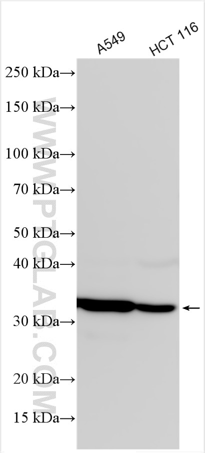
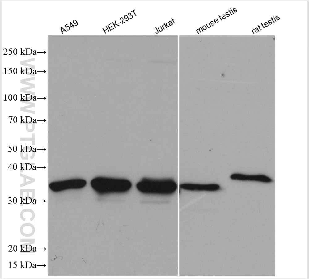
| Positive WB detected in | A549 cells, HEK-293T cells, Jurkat cells, mouse testis tissue, rat testis tissue, HCT 116 cells |
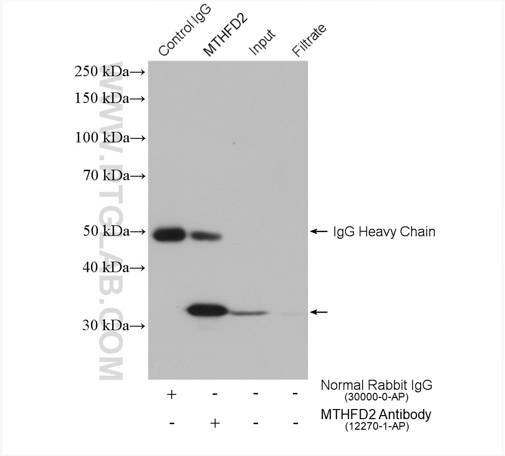
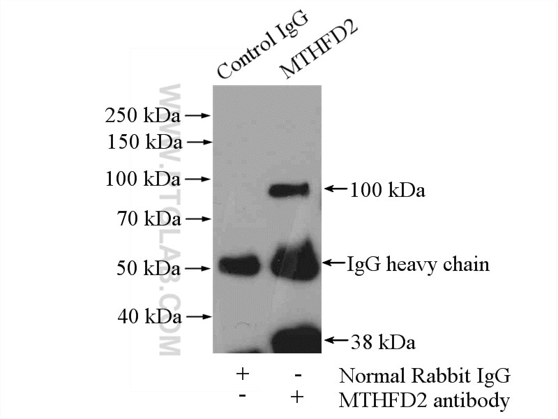
| Positive IP detected in | A549 cells, mouse testis tissue |
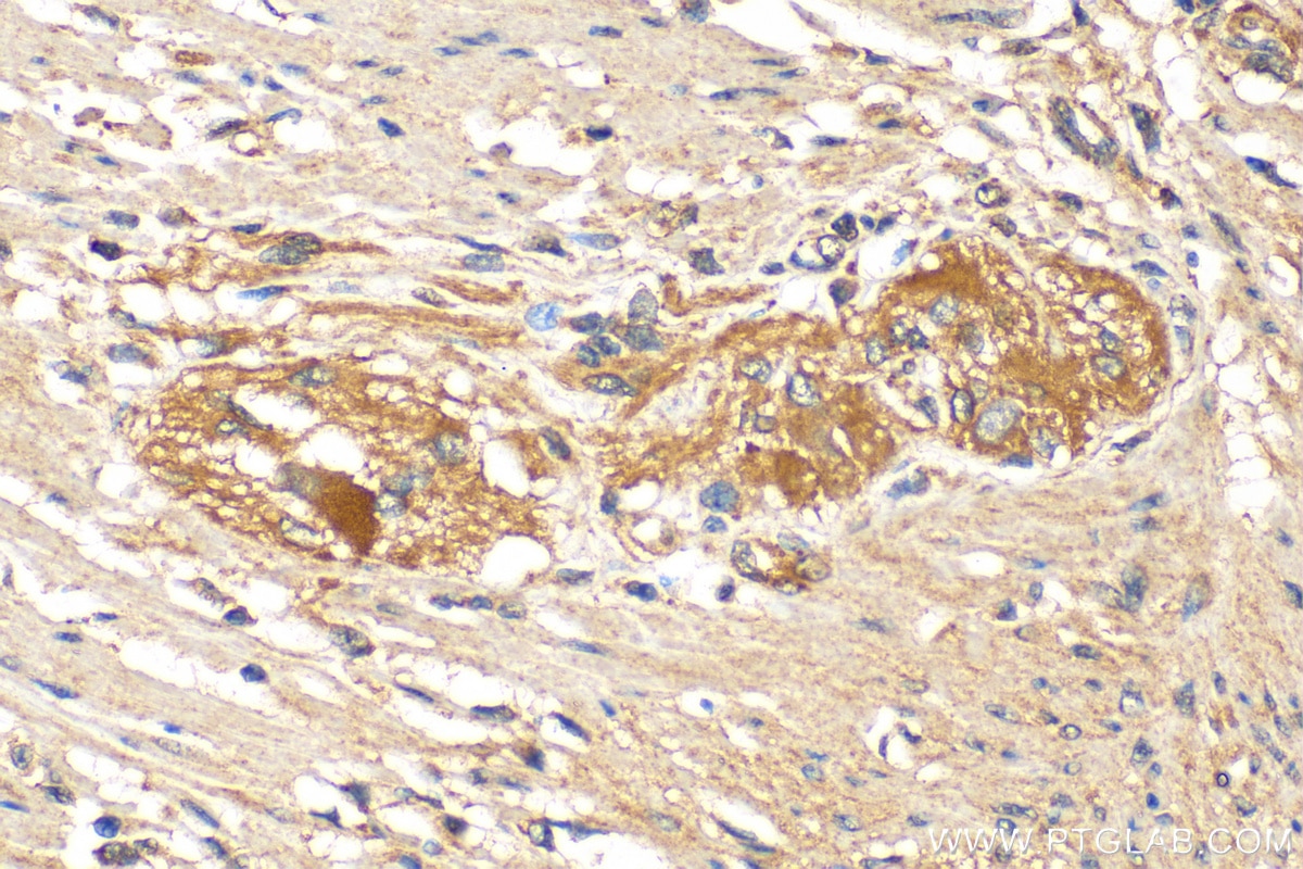
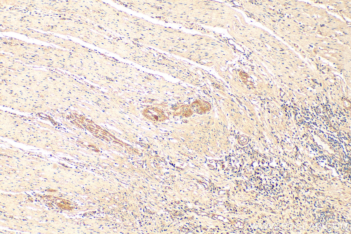
| Positive IHC detected in | human colon cancer tissue Note: suggested antigen retrieval with TE buffer pH 9.0; (*) Alternatively, antigen retrieval may be performed with citrate buffer pH 6.0 |
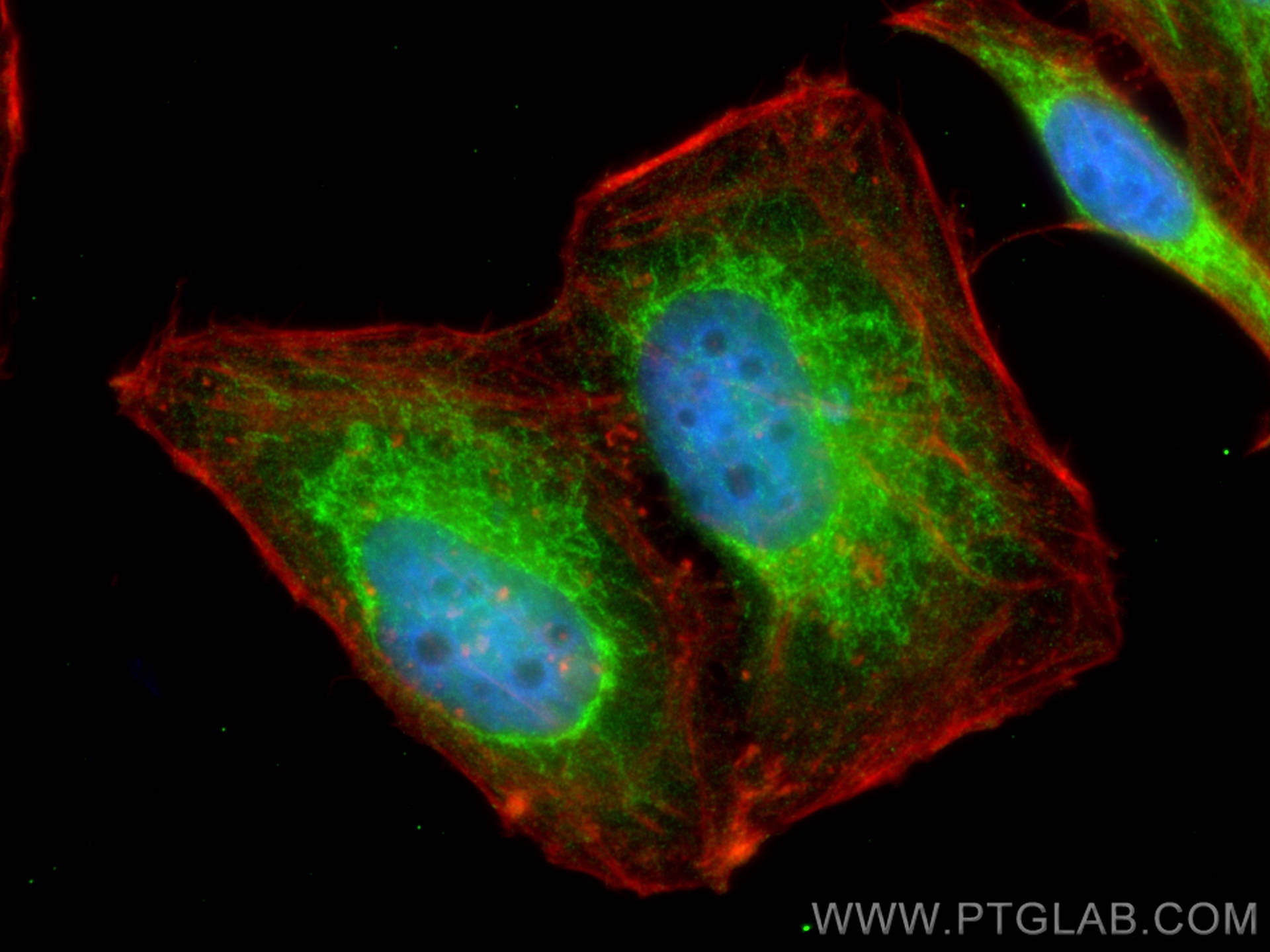
| Positive IF/ICC detected in | HepG2 cells |
Recommended dilution
| Application | Dilution |
|---|---|
| Western Blot (WB) | WB : 1:1000-1:8000 |
| Immunoprecipitation (IP) | IP : 0.5-4.0 ug for 1.0-3.0 mg of total protein lysate |
| Immunohistochemistry (IHC) | IHC : 1:200-1:800 |
| Immunofluorescence (IF)/ICC | IF/ICC : 1:200-1:800 |
| It is recommended that this reagent should be titrated in each testing system to obtain optimal results. | |
| Sample-dependent, Check data in validation data gallery. | |
Published Applications
| KD/KO | See 9 publications below |
| WB | See 47 publications below |
| IHC | See 6 publications below |
| IF | See 3 publications below |
| IP | See 2 publications below |
| CoIP | See 1 publications below |
Product Information
12270-1-AP targets MTHFD2 in WB, IHC, IF/ICC, IP, CoIP, ELISA applications and shows reactivity with human, mouse, rat samples.
| Tested Reactivity | human, mouse, rat |
| Cited Reactivity | human, mouse |
| Host / Isotype | Rabbit / IgG |
| Class | Polyclonal |
| Type | Antibody |
| Immunogen |
CatNo: Ag2911 Product name: Recombinant human MTHFD2 protein Source: e coli.-derived, PGEX-4T Tag: GST Domain: 1-350 aa of BC017054 Sequence: MAATSLMSALAARLLQPAHSCSLRLRPFHLAAVRNEAVVISGRKLAQQIKQEVRQEVEEWVASGNKRPHLSVILVGENPASHSYVLNKTRAAAVVGINSETIMKPASISEEELLNLINKLNNDDNVDGLLVQLPLPEHIDERRICNAVSPDKDVDGFHVINVGRMCLDQYSMLPATPWGVWEIIKRTGIPTLGKNVVVAGRSKNVGMPIAMLLHTDGAHERPGGDATVTISHRYTPKEQLKKHTILADIVISAAGIPNLITADMIKEGAAVIDVGINRVHDPVTAKPKLVGDVDFEGVRQKAGYITPVPGGVGPMTVAMLMKNTIIAAKKVLRLEEREVLKSKELGVATN 相同性解析による交差性が予測される生物種 |
| Full Name | methylenetetrahydrofolate dehydrogenase (NADP+ dependent) 2, methenyltetrahydrofolate cyclohydrolase |
| Calculated molecular weight | 350 aa, 38 kDa |
| Observed molecular weight | 33-38 kDa |
| GenBank accession number | BC017054 |
| Gene Symbol | MTHFD2 |
| Gene ID (NCBI) | 10797 |
| RRID | AB_2147525 |
| Conjugate | Unconjugated |
| Form | |
| Form | Liquid |
| Purification Method | Antigen affinity purification |
| UNIPROT ID | P13995 |
| Storage Buffer | PBS with 0.02% sodium azide and 50% glycerol{{ptg:BufferTemp}}7.3 |
| Storage Conditions | Store at -20°C. Stable for one year after shipment. Aliquoting is unnecessary for -20oC storage. |
Background Information
Methylenetetrahydrofolate dehydrogenase 2 (MTHFD2) is a folate-coupled mitochondrial metabolic enzyme characterized by methylenetetrahydrofolate dehydrogenase and cyclohydrolase activity. It was demonstrated that suppression of MTHFD2 inhibits methylation reactions, decreases protein synthesis and disrupts redox homeostasis, which may ultimately result in significant changes in cellular metabolic phenotype. MTHFD2 is reported to be up‐regulated in various tumours, such as lung adenocarcinoma(PMID: 34121323) and breast cancer(PMID: 34007312).
Protocols
| Product Specific Protocols | |
|---|---|
| IF protocol for MTHFD2 antibody 12270-1-AP | Download protocol |
| IHC protocol for MTHFD2 antibody 12270-1-AP | Download protocol |
| IP protocol for MTHFD2 antibody 12270-1-AP | Download protocol |
| WB protocol for MTHFD2 antibody 12270-1-AP | Download protocol |
| Standard Protocols | |
|---|---|
| Click here to view our Standard Protocols |
Publications
| Species | Application | Title |
|---|---|---|
Science mTORC1 induces purine synthesis through control of the mitochondrial tetrahydrofolate cycle. | ||
Cancer Cell Nrf2 redirects glucose and glutamine into anabolic pathways in metabolic reprogramming. | ||
Cell Metab Epstein-Barr-Virus-Induced One-Carbon Metabolism Drives B Cell Transformation. | ||
Cell Metab mTORC1 Regulates Mitochondrial Integrated Stress Response and Mitochondrial Myopathy Progression. |

