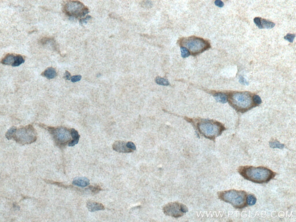
Validation Data Gallery
Tested Applications
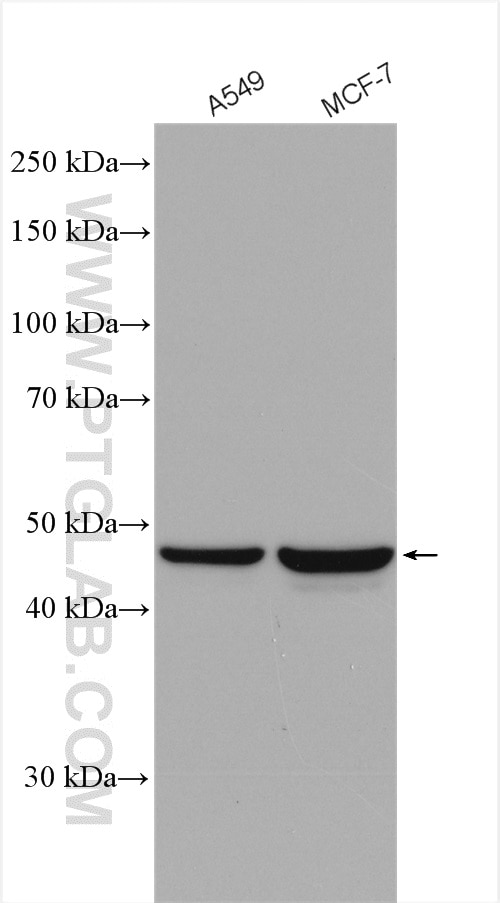
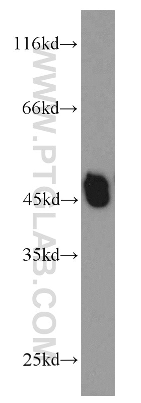
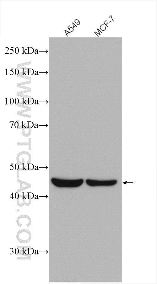
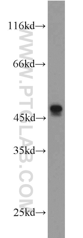
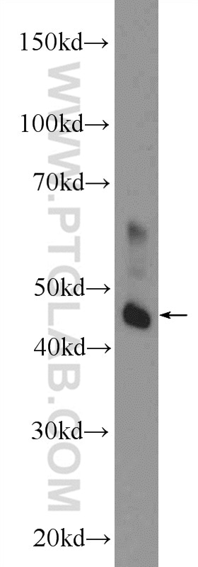
| Positive WB detected in | A549 cells, rat brain tissue, mouse small intestine tissue, SGC-7901 cells, MCF-7 cells |
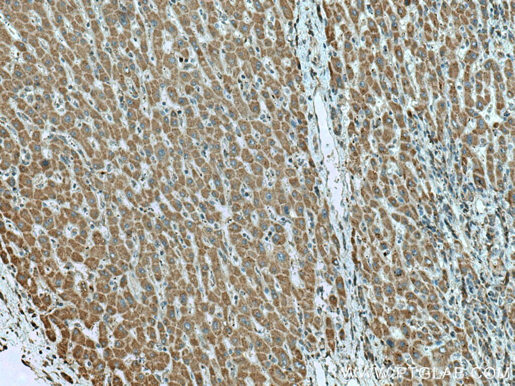
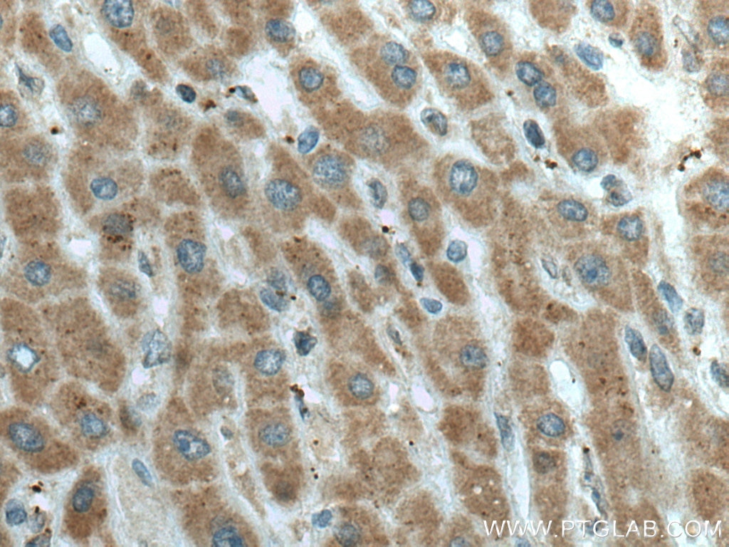
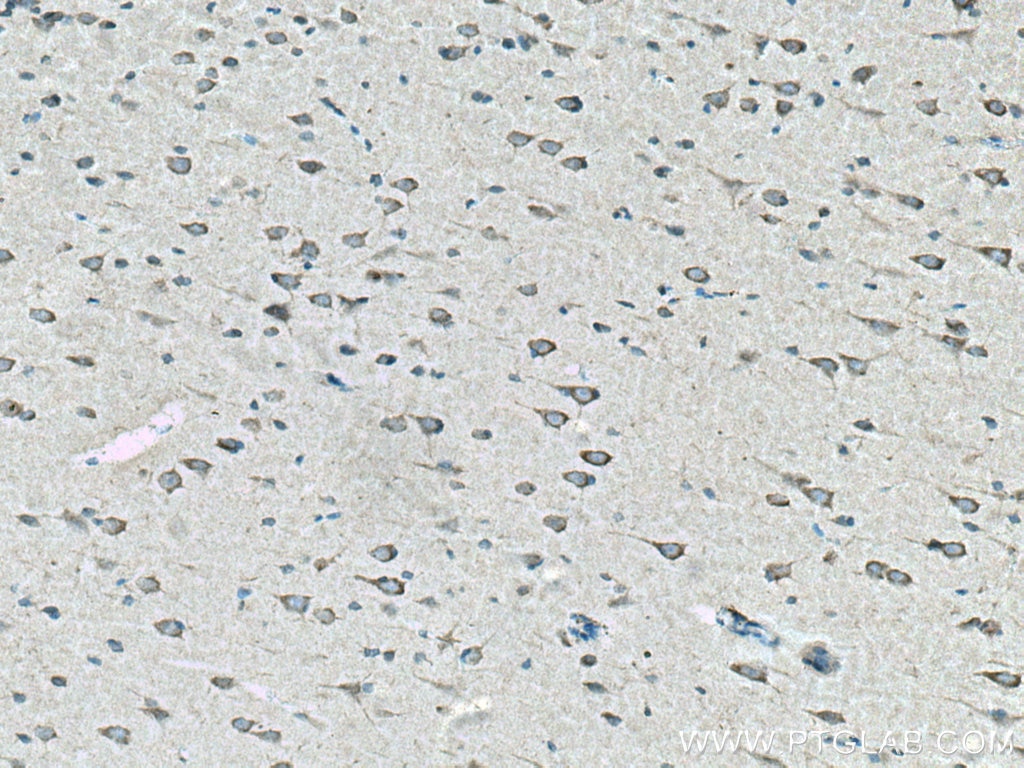
| Positive IHC detected in | human liver cancer tissue, human gliomas tissue Note: suggested antigen retrieval with TE buffer pH 9.0; (*) Alternatively, antigen retrieval may be performed with citrate buffer pH 6.0 |
Recommended dilution
| Application | Dilution |
|---|---|
| Western Blot (WB) | WB : 1:1000-1:3000 |
| Immunohistochemistry (IHC) | IHC : 1:50-1:500 |
| It is recommended that this reagent should be titrated in each testing system to obtain optimal results. | |
| Sample-dependent, Check data in validation data gallery. | |
Published Applications
| KD/KO | See 7 publications below |
| WB | See 15 publications below |
| IHC | See 1 publications below |
Product Information
15707-1-AP targets L2HGDH in WB, IHC, ELISA applications and shows reactivity with human, mouse, rat samples.
| Tested Reactivity | human, mouse, rat |
| Cited Reactivity | human, mouse |
| Host / Isotype | Rabbit / IgG |
| Class | Polyclonal |
| Type | Antibody |
| Immunogen |
CatNo: Ag8382 Product name: Recombinant human L2HGDH protein Source: e coli.-derived, PET28a Tag: 6*His Domain: 1-351 aa of BC006117 Sequence: MVPALRYLVGACGRARGRFAGGSPGACGFASGRPRPLCGGSRSASTSSFDIVIVGGGIVGLASARALILRHPSLSIGVLEKEKDLAVHQTGHNSGVIHSGIYYKPESLKAKLCVQGAALLYEYCQQKGISYKQCGKLIVAVEQEEIPRLQALYEKGLQNGVPGLRLIQQEDIKKKEPYCRGLMAIDCPHTGIVDYRQVALSFAQDFQEAGGSVLTNFEVKGIEMAKESPSRSIDGMQYPIVIKNTKGEEIRCQYVVTCAGLYSDRISELSGCTPDPRIVPFRGDYLLLKPEKCYLVKGNIYPVPDSRFPFLGVHFTPRMDGSIWLGPNAVLAFKREGYRPFDFSATDVMDI 相同性解析による交差性が予測される生物種 |
| Full Name | L-2-hydroxyglutarate dehydrogenase |
| Calculated molecular weight | 463aa,50 kDa; 441aa,49 kDa |
| Observed molecular weight | 45 kDa |
| GenBank accession number | BC006117 |
| Gene Symbol | L2HGDH |
| Gene ID (NCBI) | 79944 |
| RRID | AB_2133202 |
| Conjugate | Unconjugated |
| Form | |
| Form | Liquid |
| Purification Method | Antigen affinity purification |
| UNIPROT ID | Q9H9P8 |
| Storage Buffer | PBS with 0.02% sodium azide and 50% glycerol{{ptg:BufferTemp}}7.3 |
| Storage Conditions | Store at -20°C. Stable for one year after shipment. Aliquoting is unnecessary for -20oC storage. |
Background Information
L2HGDH(L-2-hydroxyglutarate dehydrogenase, mitochondrial) is also named as duranin, C14orf160 and belongs to the L2HGDH family. The putative L2HGDH is predicted to be targeted to the mitochondria where its mitochondrial targeting sequence is presumably removed(PMID:16005139). Defects in L2HGDH are the cause of L-2-hydroxyglutaric aciduria (L2HGA). It has 2 isoforms produced by alternative splicing with the molecular weight of 50 kDa and 48 kDa. L2HGDH also can be detected as ~45kD due to the 51aa transit peptide cleaved.
Protocols
| Product Specific Protocols | |
|---|---|
| IHC protocol for L2HGDH antibody 15707-1-AP | Download protocol |
| WB protocol for L2HGDH antibody 15707-1-AP | Download protocol |
| Standard Protocols | |
|---|---|
| Click here to view our Standard Protocols |
Publications
| Species | Application | Title |
|---|---|---|
Cell Metab Hypoxia-Mediated Increases in L-2-hydroxyglutarate Coordinate the Metabolic Response to Reductive Stress.
| ||
Sci Transl Med 2-Hydroxyglutarate produced by neomorphic IDH mutations suppresses homologous recombination and induces PARP inhibitor sensitivity.
| ||
Sci Adv Lipoylation inhibition enhances radiation control of lung cancer by suppressing homologous recombination DNA damage repair | ||
Cancer Discov L-2-Hydroxyglutarate: an epigenetic modifier and putative oncometabolite in renal cancer. | ||
Cell Chem Biol MYC Regulation of D2HGDH and L2HGDH Influences the Epigenome and Epitranscriptome.
|

