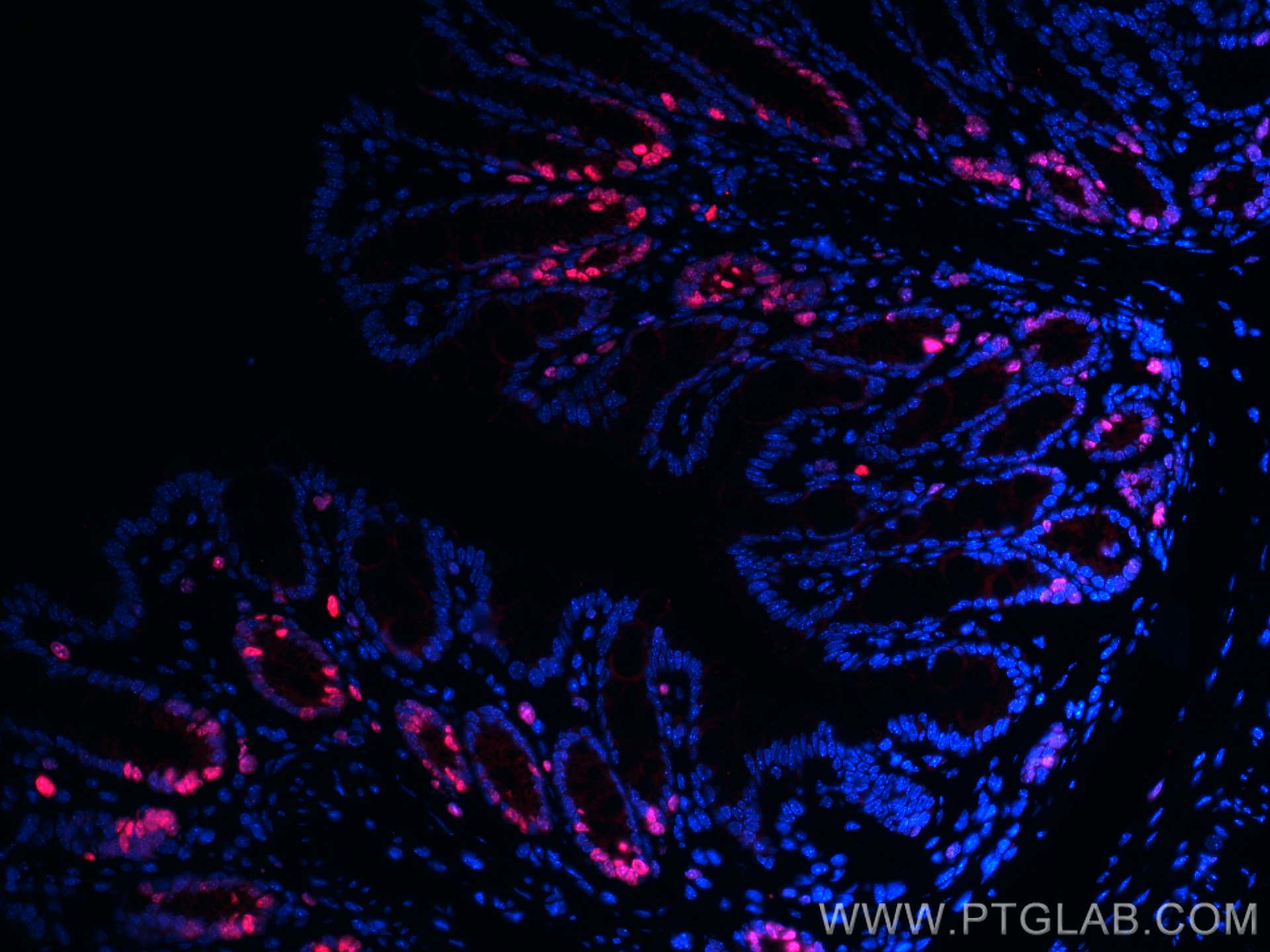
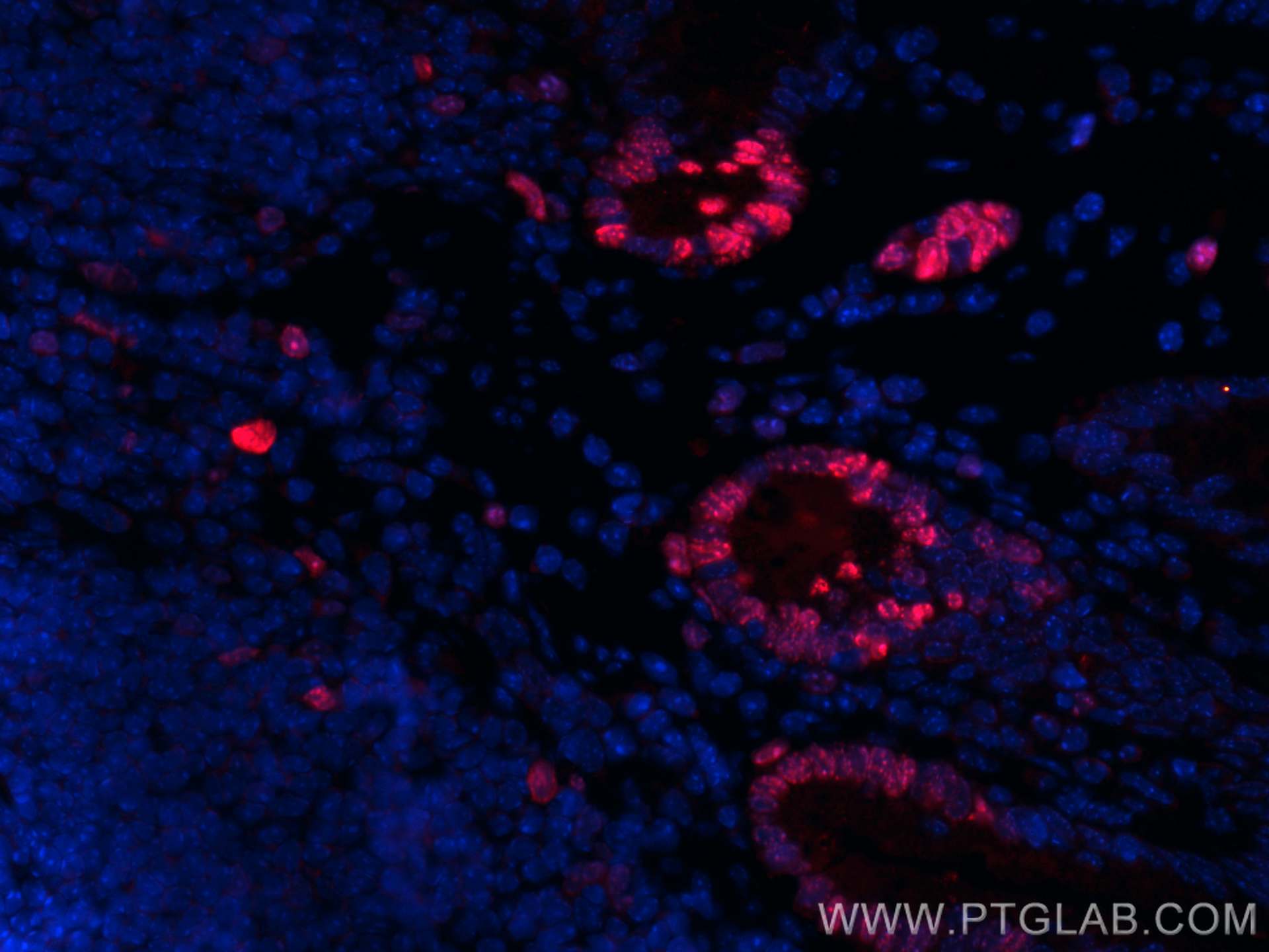
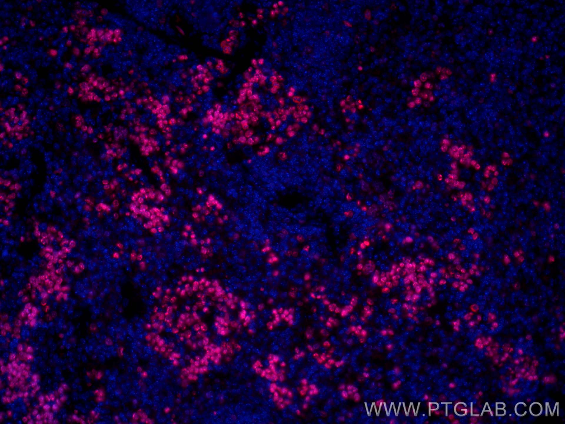
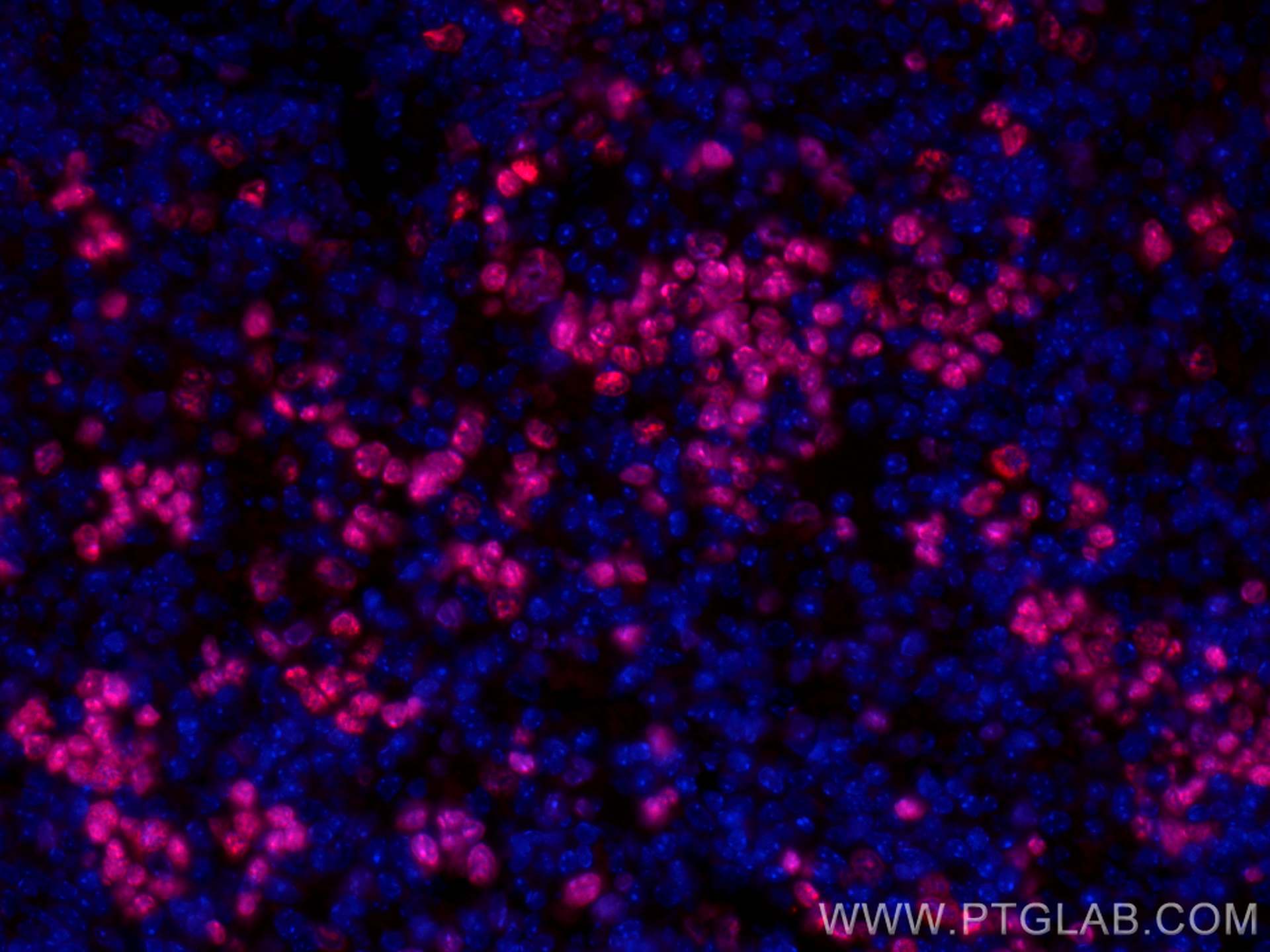
CoraLite® Plus 594-conjugated Ki-67 Polyclonal antibody
Ki-67 Polyclonal Antibody for IF-P
Host / Isotype
Rabbit / IgG
Reactivity
mouse
Applications
IF-P
Conjugate
CoraLite® Plus 594 Fluorescent Dye
Cat no : CL594-28074
Synonyms
Validation Data Gallery
Tested Applications
| Positive IF-P detected in | mouse colon tissue, mouse spleen tissue |
Recommended dilution
| Application | Dilution |
|---|---|
| Immunofluorescence (IF)-P | IF-P : 1:50-1:500 |
| It is recommended that this reagent should be titrated in each testing system to obtain optimal results. | |
| Sample-dependent, Check data in validation data gallery. | |
Product Information
CL594-28074 targets Ki-67 in IF-P applications and shows reactivity with mouse samples.
| Tested Reactivity | mouse |
| Host / Isotype | Rabbit / IgG |
| Class | Polyclonal |
| Type | Antibody |
| Immunogen | Ki-67 fusion protein Ag27894 相同性解析による交差性が予測される生物種 |
| Full Name | antigen identified by monoclonal antibody Ki 67 |
| Calculated molecular weight | 351 kDa |
| GenBank accession number | NM_001081117 |
| Gene symbol | ki67 |
| Gene ID (NCBI) | 17345 |
| Conjugate | CoraLite® Plus 594 Fluorescent Dye |
| Excitation/Emission maxima wavelengths | 594 nm / 615 nm |
| Form | Liquid |
| Purification Method | Antigen affinity purification |
| Storage Buffer | PBS with 50% Glycerol, 0.05% Proclin300, 0.5% BSA, pH 7.3. |
| Storage Conditions | Store at -20°C. Avoid exposure to light. Stable for one year after shipment. Aliquoting is unnecessary for -20oC storage. |
Background Information
The Ki-67 protein (also known as MKI67) is a cellular marker for proliferation. Ki67 is present during all active phases of the cell cycle (G1, S, G2 and M), but is absent in resting cells (G0). Cellular content of Ki-67 protein markedly increases during cell progression through S phase of the cell cycle. Therefore, the nuclear expression of Ki67 can be evaluated to assess tumor proliferation by immunohistochemistry.
Protocols
| Product Specific Protocols | |
|---|---|
| IF protocol for CL Plus 594 Ki-67 antibody CL594-28074 | Download protocol |
| Standard Protocols | |
|---|---|
| Click here to view our Standard Protocols |


