CoraLite® Plus 488-conjugated PD-1/CD279 Monoclonal antibody
PD-1/CD279 Monoclonal Antibody for FC, IF
Host / Isotype
Mouse / IgG2b
Reactivity
human, rat, mouse
Applications
IF, FC
Conjugate
CoraLite® Plus 488 Fluorescent Dye
CloneNo.
4H4D1
Cat no : CL488-66220
Synonyms
Validation Data Gallery
Tested Applications
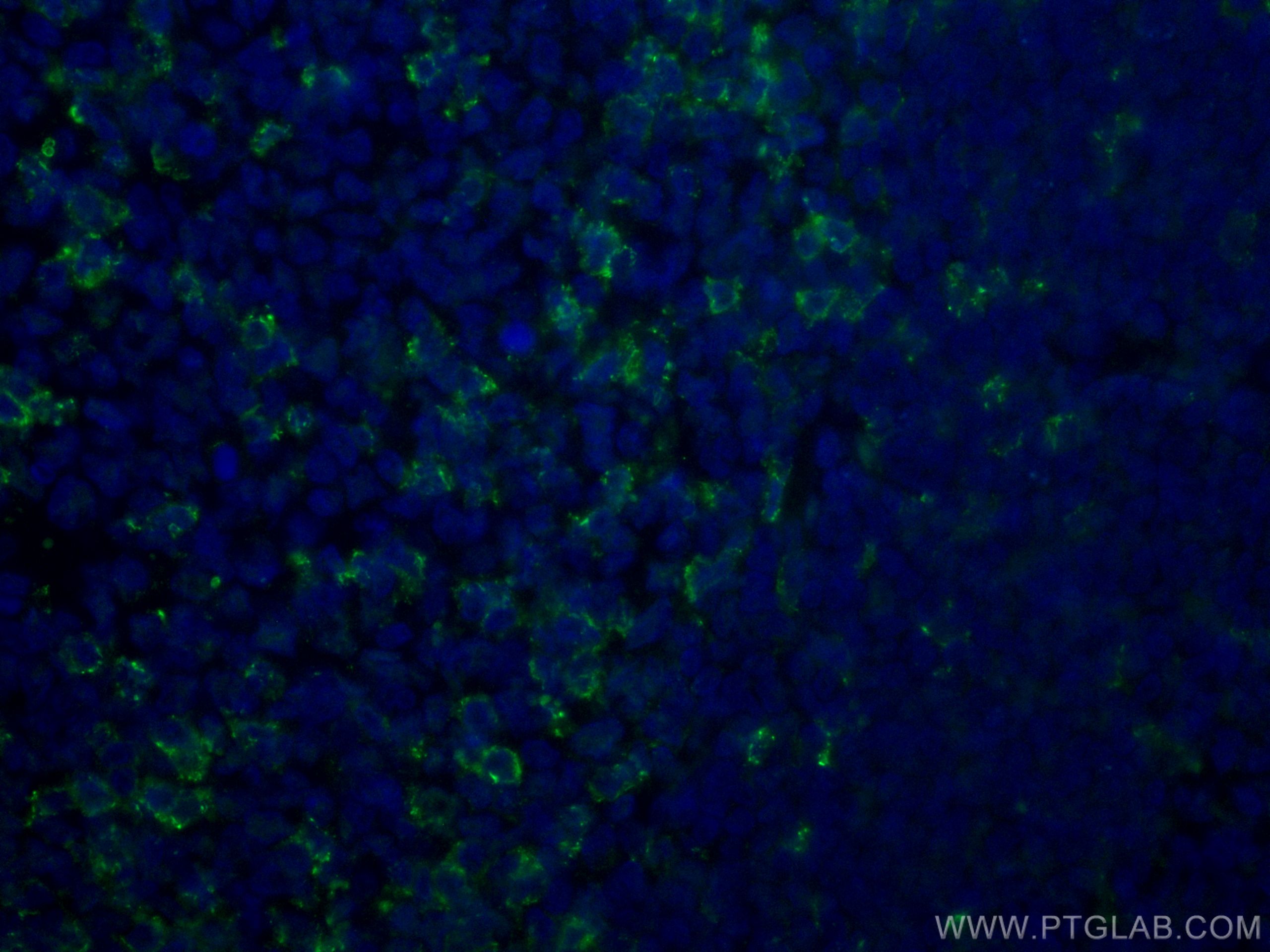
| Positive IF detected in | human tonsillitis tissue |
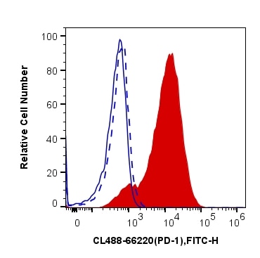
| Positive FC detected in | CD3 antibody treated Jurkat cells |
Recommended dilution
| Application | Dilution |
|---|---|
| Immunofluorescence (IF) | IF : 1:50-1:500 |
| Flow Cytometry (FC) | FC : 0.40 ug per 10^6 cells in a 100 µl suspension |
| Sample-dependent, check data in validation data gallery | |
Product Information
CL488-66220 targets PD-1/CD279 in IF, FC applications and shows reactivity with human, rat, mouse samples.
| Tested Reactivity | human, rat, mouse |
| Host / Isotype | Mouse / IgG2b |
| Class | Monoclonal |
| Type | Antibody |
| Immunogen | PD-1/CD279 fusion protein Ag12470 相同性解析による交差性が予測される生物種 |
| Full Name | programmed cell death 1 |
| Calculated molecular weight | 288 aa, 32 kDa |
| GenBank accession number | BC074740 |
| Gene symbol | PDCD1 |
| Gene ID (NCBI) | 5133 |
| RRID | AB_2883287 |
| Conjugate | CoraLite® Plus 488 Fluorescent Dye |
| Excitation/Emission maxima wavelengths | 493 nm / 522 nm |
| Form | Liquid |
| Purification Method | Protein A purification |
| Storage Buffer | PBS with 50% Glycerol, 0.05% Proclin300, 0.5% BSA, pH 7.3. |
| Storage Conditions | Store at -20°C. Avoid exposure to light. Stable for one year after shipment. Aliquoting is unnecessary for -20oC storage. |
Background Information
Programmed cell death 1 (PD-1, also known as CD279) is an immunoinhibitory receptor that belongs to the CD28/CTLA-4 subfamily of the Ig superfamily. It is a 288 amino acid (aa) type I transmembrane protein composed of one Ig superfamily domain, a stalk, a transmembrane domain, and an intracellular domain containing an immunoreceptor tyrosine-based inhibitory motif (ITIM) as well as an immunoreceptor tyrosine-based switch motif (ITSM) (PMID: 18173375). PD-1 is expressed during thymic development and is induced in a variety of hematopoietic cells in the periphery by antigen receptor signaling and cytokines (PMID: 20636820). Engagement of PD-1 by its ligands PD-L1 or PD-L2 transduces a signal that inhibits T-cell proliferation, cytokine production, and cytolytic function (PMID: 19426218). It is critical for the regulation of T cell function during immunity and tolerance. Blockade of PD-1 can overcome immune resistance and also has been shown to have antitumor activity (PMID: 22658127; 23169436). The calculated molecular weight of PD-1 is 32 kDa. It has been reported that PD-1 is heavily glycosylated and migrates with an apparent molecular mass of 47-55 kDa on SDS-PAGE (PMID: 8671665; 17640856; 17003438). This anitbody is CL488(Ex/Em 488 nm/515 nm) conjugated.
Protocols
| Product Specific Protocols | |
|---|---|
| IF protocol for CL Plus 488 PD-1/CD279 antibody CL488-66220 | Download protocol |
| Standard Protocols | |
|---|---|
| Click here to view our Standard Protocols |


