MAOB Monoclonal antibody
MAOB Monoclonal Antibody for IF, IHC, WB, ELISA
Host / Isotype
Mouse / IgG1
Reactivity
human, mouse
Applications
WB, IHC, IF, ELISA
Conjugate
Unconjugated
CloneNo.
2B12F3
Cat no : 66107-1-Ig
Synonyms
Validation Data Gallery
Tested Applications
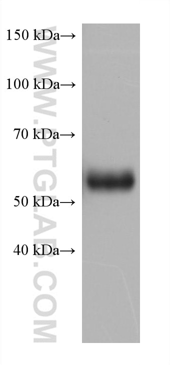
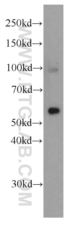
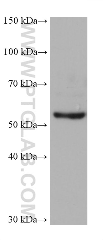
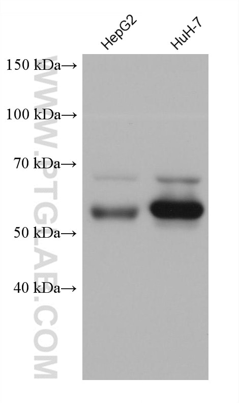
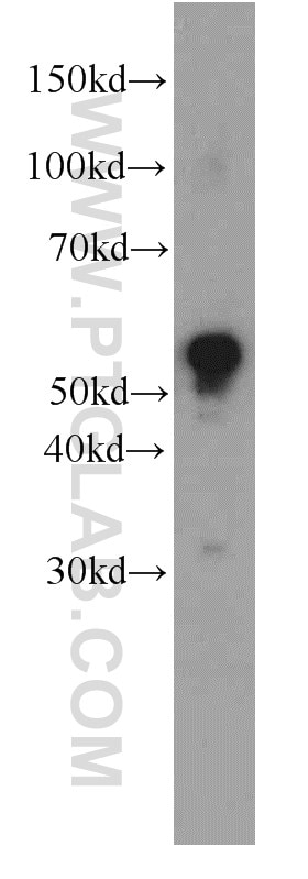
| Positive WB detected in | fetal human brain tissue, human brain tissue, human kidney tissue, HeLa cells, HepG2 cells, HuH-7 cells |
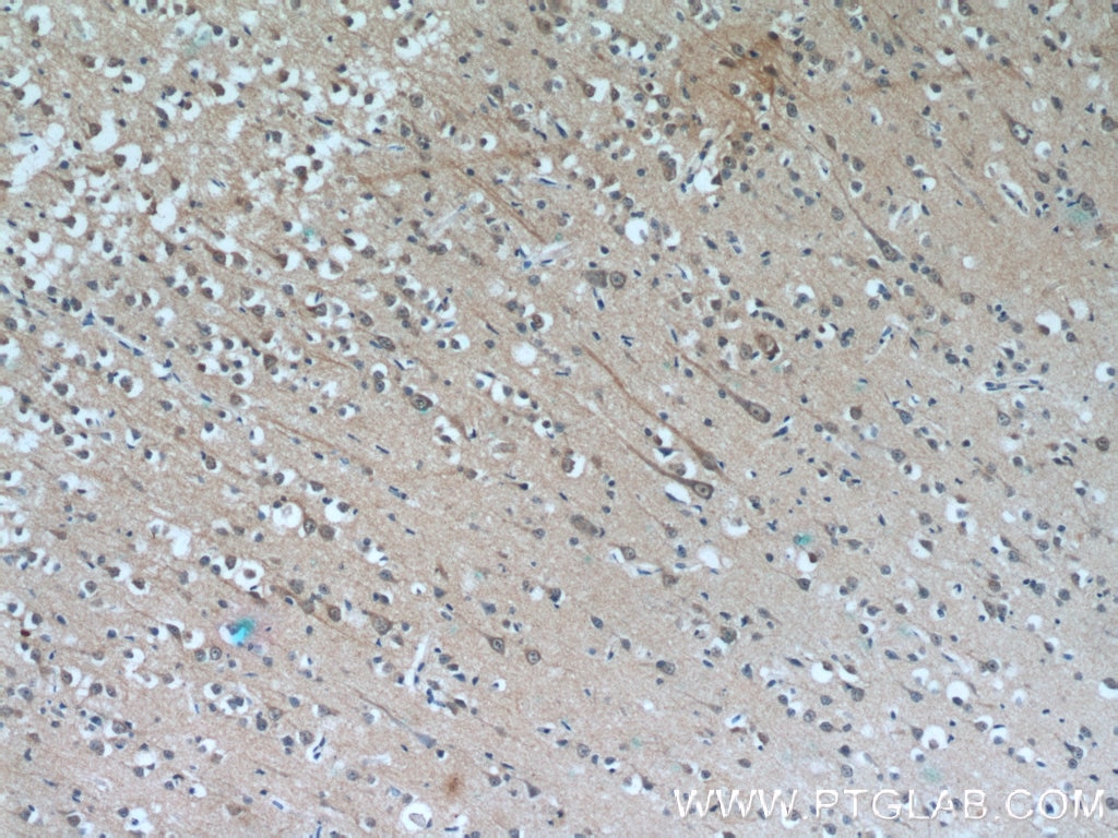
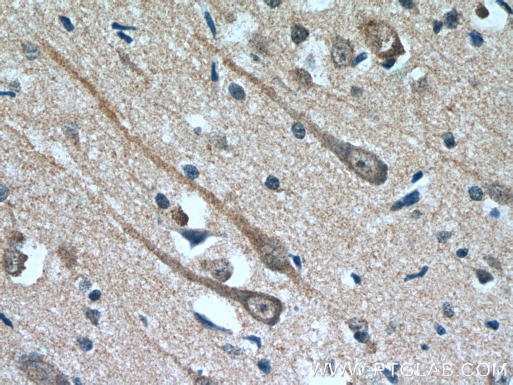
| Positive IHC detected in | human brain tissue Note: suggested antigen retrieval with TE buffer pH 9.0; (*) Alternatively, antigen retrieval may be performed with citrate buffer pH 6.0 |
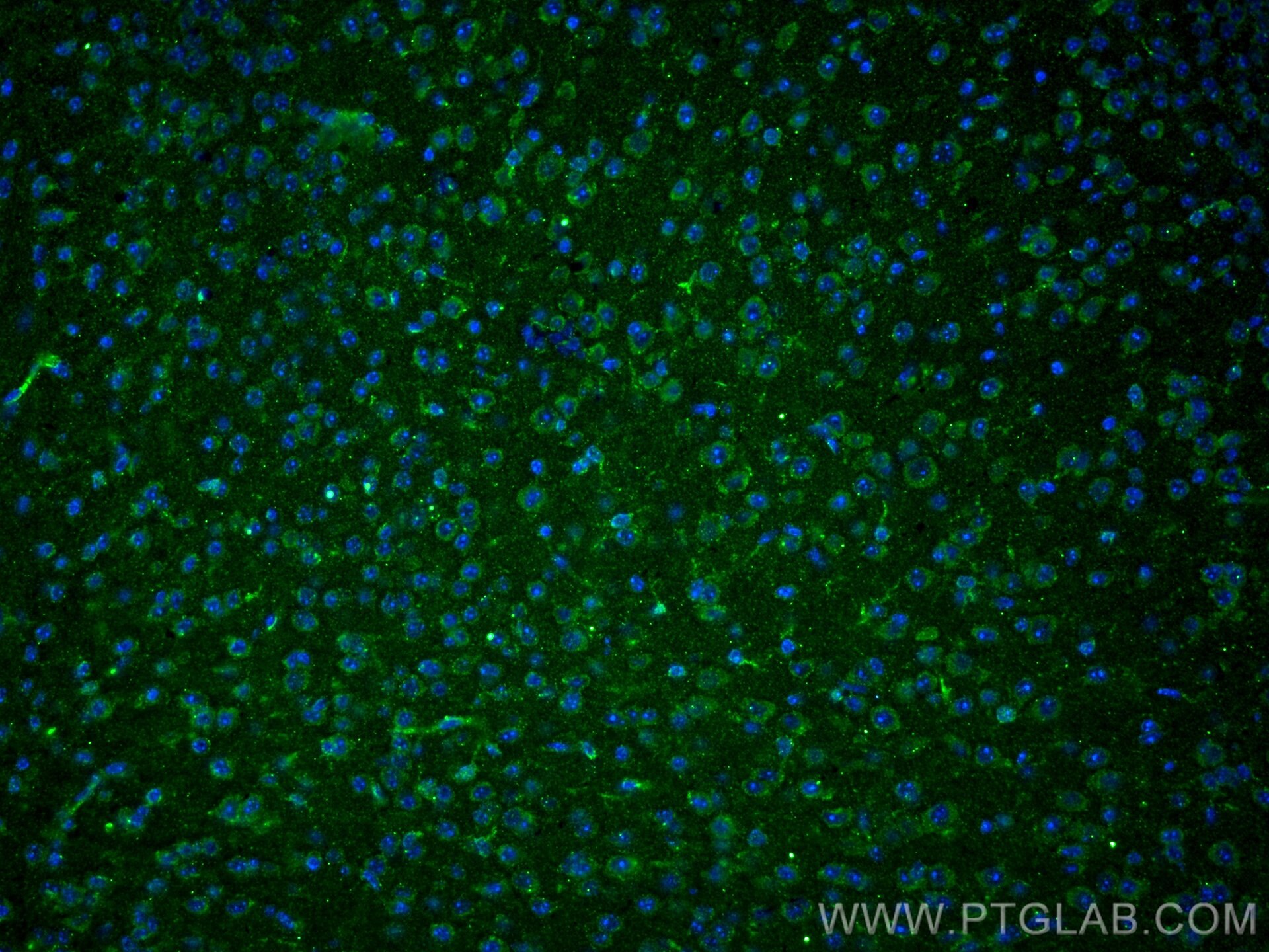
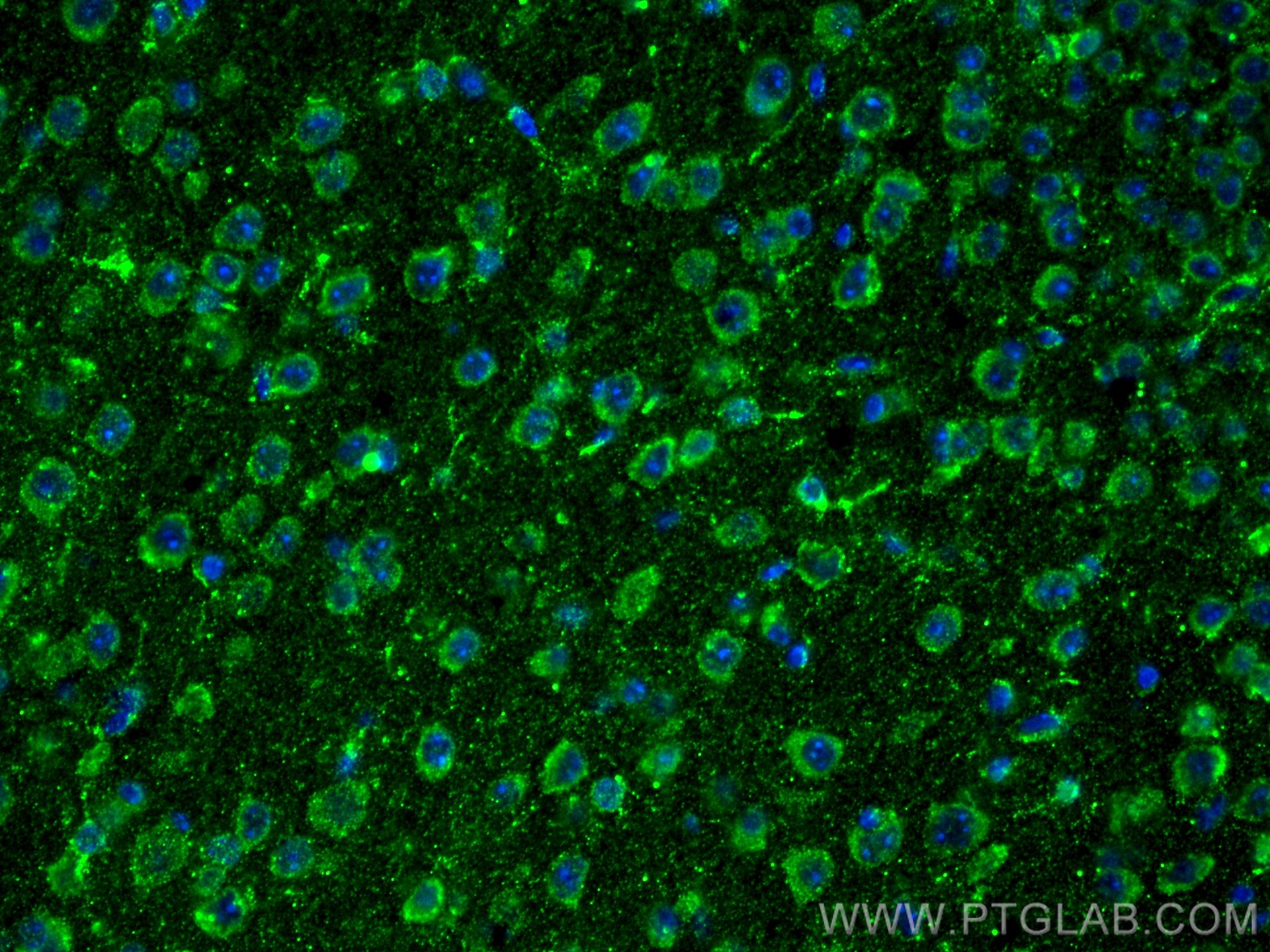
| Positive IF detected in | mouse brain tissue |
Recommended dilution
| Application | Dilution |
|---|---|
| Western Blot (WB) | WB : 1:2000-1:10000 |
| Immunohistochemistry (IHC) | IHC : 1:20-1:200 |
| Immunofluorescence (IF) | IF : 1:10-1:100 |
| Sample-dependent, check data in validation data gallery | |
Product Information
66107-1-Ig targets MAOB in WB, IHC, IF, ELISA applications and shows reactivity with human, mouse samples.
| Tested Reactivity | human, mouse |
| Host / Isotype | Mouse / IgG1 |
| Class | Monoclonal |
| Type | Antibody |
| Immunogen | MAOB fusion protein Ag17918 相同性解析による交差性が予測される生物種 |
| Full Name | monoamine oxidase B |
| Calculated molecular weight | 520 aa, 59 kDa |
| Observed molecular weight | 59 kDa |
| GenBank accession number | BC022494 |
| Gene symbol | MAOB |
| Gene ID (NCBI) | 4129 |
| RRID | AB_2881506 |
| Conjugate | Unconjugated |
| Form | Liquid |
| Purification Method | Protein A purification |
| Storage Buffer | PBS with 0.02% sodium azide and 50% glycerol pH 7.3. |
| Storage Conditions | Store at -20°C. Stable for one year after shipment. Aliquoting is unnecessary for -20oC storage. |
Background Information
The MAOB gene encodes a 520-amino acid protein with a molecular mass of 58.8 kD and shows 70% amino acid identity to MAOA. MAOA and MAOB are present in the outer mitochondrial membrane in the central nervous system and peripheral tissues.A comparison of highly purified human placental MAOA and human liver MAOB revealed that the A form of the enzyme is larger by 2 kDa and only one potential N-glycosylation site exists in each protein, with each site in a different relevant position(PMID:3387449).
Protocols
| Product Specific Protocols | |
|---|---|
| WB protocol for MAOB antibody 66107-1-Ig | Download protocol |
| IHC protocol for MAOB antibody 66107-1-Ig | Download protocol |
| IF protocol for MAOB antibody 66107-1-Ig | Download protocol |
| Standard Protocols | |
|---|---|
| Click here to view our Standard Protocols |
