- Featured Product
- KD/KO Validated
G3BP1 Monoclonal antibody
G3BP1 Monoclonal Antibody for IF, IHC, WB, ELISA
Host / Isotype
Mouse / IgG1
Reactivity
Human, mouse, rat, pig and More (1)
Applications
WB, RIP, IP, IHC, IF, FC, CoIP, ELISA
Conjugate
Unconjugated
CloneNo.
1E4A2
Cat no : 66486-1-Ig
Synonyms
Validation Data Gallery
 at dilution of 1:20000 incubated at room temperature for 1.5 hours.")
 at dilution of 1:40000 incubated at room temperature for 1.5 hours. The membrane was stripped and reblotted with HRP-conjugated GAPDH Monoclonal antibody (<a class='green' href='/productredirect?CatalogNo=HRP-60004' target='_blank'>HRP-60004</a>) as loading control.")
 at dilution of 1:20000 incubated at room temperature for 1.5 hours.")
 at dilution of 1:10000 incubated at room temperature for 1.5 hours.")
 at dilution of 1:10000 incubated at room temperature for 1.5 hours.")
 at dilution of 1:200 (under 10x lens).")
 at dilution of 1:200 (under 40x lens).")
 at dilution of 1:200 (under 10x lens. Heat mediated antigen retrieval with Tris-EDTA buffer (pH 9.0).")
 at dilution of 1:200 (under 40x lens. Heat mediated antigen retrieval with Tris-EDTA buffer (pH 9.0).")
 at dilution of 1:200 (under 10x lens).")
 at dilution of 1:200 (under 40x lens).")
 at dilution of 1:5000 (under 10x lens). Heat mediated antigen retrieval with Tris-EDTA buffer (pH 9.0).")
 at dilution of 1:5000 (under 40x lens). Heat mediated antigen retrieval with Tris-EDTA buffer (pH 9.0).")
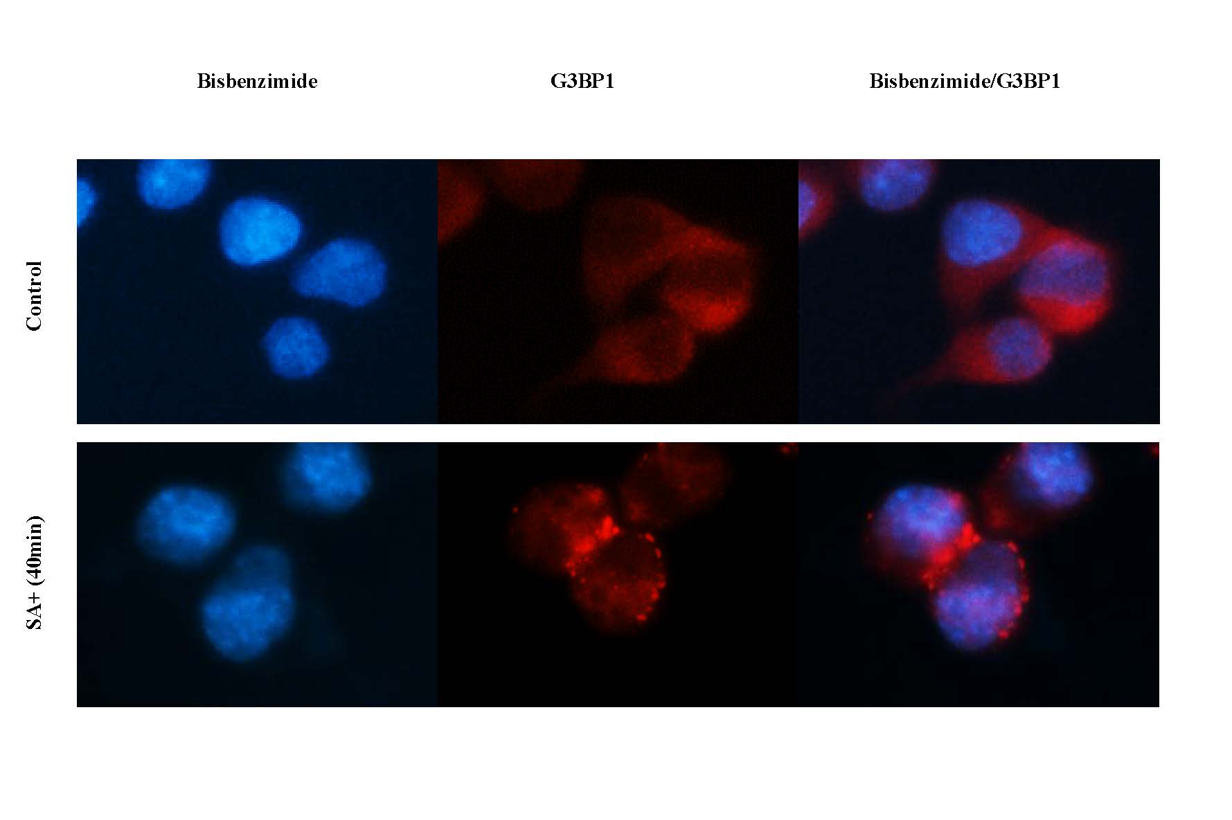
 fixed sodium arsenite treated HeLa cells using <a class='green' href='/productredirect?CatalogNo=G3' target='_blank'>G3</a><a class='green' href='/productredirect?CatalogNo=BP1' target='_blank'>BP1</a> antibody (<a class='green' href='/productredirect?CatalogNo=66486-1-Ig' target='_blank'>66486-1-Ig</a>, Clone: 1<a class='green' href='/productredirect?CatalogNo=E4' target='_blank'>E4</a><a class='green' href='/productredirect?CatalogNo=A2' target='_blank'>A2</a> ) at dilution of 1:1000 and CoraLite®488-Conjugated AffiniPure Goat Anti-Mouse IgG(H+L), <a class='green' href='/productredirect?CatalogNo=CL59' target='_blank'>CL59</a>4-Phalloidin (red).")
Tested Applications
| Positive WB detected in | LNCaP cells, RAW 264.7 cells, pig brain tissue, HeLa cells, mouse brain tissue, HEK-293 cells, HepG2 cells, Jurkat cells, K-562 cells, HSC-T6 cells, PC-12 cells, NIH/3T3 cells, 4T1 cells |
| Positive IHC detected in | human testis tissue, human colon tissue, human lymphoma tissue, rat brain tissue Note: suggested antigen retrieval with TE buffer pH 9.0; (*) Alternatively, antigen retrieval may be performed with citrate buffer pH 6.0 |
| Positive IF detected in | sodium arsenite treated HeLa cells |
Recommended dilution
| Application | Dilution |
|---|---|
| Western Blot (WB) | WB : 1:5000-1:50000 |
| Immunohistochemistry (IHC) | IHC : 1:50-1:500 |
| Immunofluorescence (IF) | IF : 1:500-1:2000 |
| Sample-dependent, check data in validation data gallery | |
Product Information
66486-1-Ig targets G3BP1 in WB, RIP, IP, IHC, IF, FC, CoIP, ELISA applications and shows reactivity with Human, mouse, rat, pig samples.
| Tested Reactivity | Human, mouse, rat, pig |
| Cited Reactivity | human, rat, mouse |
| Host / Isotype | Mouse / IgG1 |
| Class | Monoclonal |
| Type | Antibody |
| Immunogen | G3BP1 fusion protein Ag3728 相同性解析による交差性が予測される生物種 |
| Full Name | GTPase activating protein (SH3 domain) binding protein 1 |
| Calculated molecular weight | 466 aa, 52 kDa |
| Observed molecular weight | 68 kDa |
| GenBank accession number | BC006997 |
| Gene symbol | G3BP1 |
| Gene ID (NCBI) | 10146 |
| RRID | AB_2819031 |
| Conjugate | Unconjugated |
| Form | Liquid |
| Purification Method | Protein A purification |
| Storage Buffer | PBS with 0.02% sodium azide and 50% glycerol pH 7.3. |
| Storage Conditions | Store at -20°C. Stable for one year after shipment. Aliquoting is unnecessary for -20oC storage. |
Background Information
GAP SH3 Binding Protein 1 (G3BP1), also named as G3BP, is an effector of stress granule (SG) assembly. SG biology plays an important role in the pathophysiology of TDP-43 in ALS and FTLD-U. G3BP1 can be used as a marker of SG. It has been shown to function downstream of Ras and play a role in RNA metabolism, signal transduction, and proliferation. G3BP1 is a ubiquitously expressed protein that localizes to the cytoplasm in proliferating cells and to the nucleus in non-proliferating cells. G3BP1 has recently been implicated in cancer biology.
Protocols
| Product Specific Protocols | |
|---|---|
| WB protocol for G3BP1 antibody 66486-1-Ig | Download protocol |
| IHC protocol for G3BP1 antibody 66486-1-Ig | Download protocol |
| IF protocol for G3BP1 antibody 66486-1-Ig | Download protocol |
| Standard Protocols | |
|---|---|
| Click here to view our Standard Protocols |
Publications
| Species | Application | Title |
|---|---|---|
Nature DDX3X acts as a live-or-die checkpoint in stressed cells by regulating NLRP3 inflammasome. | ||
Sci Transl Med Precise genomic editing of pathogenic mutations in RBM20 rescues dilated cardiomyopathy | ||
Nat Struct Mol Biol TDP-43 aggregation induced by oxidative stress causes global mitochondrial imbalance in ALS. | ||
Autophagy Stress granule homeostasis is modulated by TRIM21-mediated ubiquitination of G3BP1 and autophagy-dependent elimination of stress granules | ||
Nat Chem Biol K29-linked ubiquitin signaling regulates proteotoxic stress response and cell cycle. |
Reviews
The reviews below have been submitted by verified Proteintech customers who received an incentive forproviding their feedback.
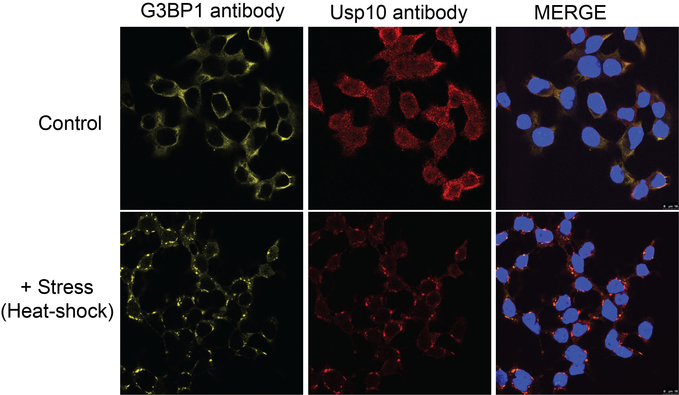
FH Haibo (Verified Customer) (10-26-2020) | This is a excellent antibody to visualize stress granules tested in HEK293 and human fibroblasts.
|
FH Manohar (Verified Customer) (09-23-2020) | 1% milk is used
|
FH David (Verified Customer) (01-14-2020) | Good signal in control cells and immunopositive puncta seen in response to stress (e.g. sodium arsenite).
|
FH Azita (Verified Customer) (10-04-2019) | Assembly of stress granula upon treatment with sodium arsenite for 40 min. (It works great)
 |
FH Kyosuke (Verified Customer) (06-12-2019) | It works well on HEK293T for Western blot.
|
FH Natalia (Verified Customer) (06-06-2019) | PFA fixated cells
 |

