- Featured Product
- KD/KO Validated
CCR2a-specific Polyclonal antibody
CCR2a-specific Polyclonal Antibody for FC, IHC, WB, ELISA
Host / Isotype
Rabbit / IgG
Reactivity
human, mouse, rat
Applications
WB, IHC, IF, FC, ELISA
Conjugate
Unconjugated
Cat no : 16153-1-AP
Synonyms
Validation Data Gallery
 with sh-Control and sh-<a class='green' href='/productredirect?CatalogNo=CCR2' target='_blank'>CCR2</a>a-specific transfected Jurkat cells.")
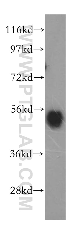
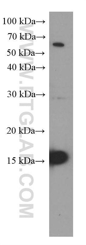
 at dilution of 1:2000 incubated at room temperature for 1.5 hours.")
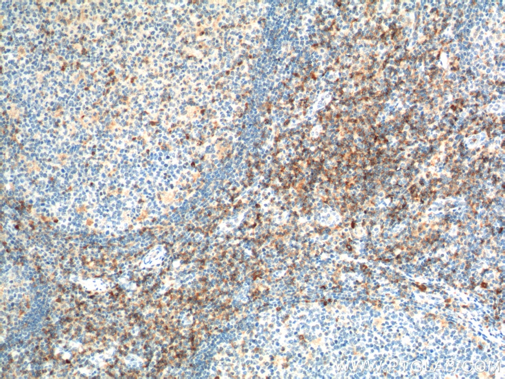
 at dilution of 1:200 (under 40x lens). Heat mediated antigen retrieval with Tris-EDTA buffer (pH 9.0).")


 and control antibody (blue). Fixed with 90% MeOH blocked with 3% BSA (30 min). Alexa Fluor 488-conjugated AffiniPure Goat Anti-Rabbit IgG(H+L) with dilution 1:1000.")
Tested Applications
| Positive WB detected in | Jurkat cells, mouse skeletal muscle tissue, rat skeletal muscle tissue |
| Positive IHC detected in | mouse kidney tissue, human kidney tissue, human ovary tumor tissue Note: suggested antigen retrieval with TE buffer pH 9.0; (*) Alternatively, antigen retrieval may be performed with citrate buffer pH 6.0 |
| Positive FC detected in | Jurkat cells |
Recommended dilution
| Application | Dilution |
|---|---|
| Western Blot (WB) | WB : 1:1000-1:4000 |
| Immunohistochemistry (IHC) | IHC : 1:50-1:500 |
| Flow Cytometry (FC) | FC : 0.20 ug per 10^6 cells in a 100 µl suspension |
| Sample-dependent, check data in validation data gallery | |
Published Applications
| WB | See 11 publications below |
| IHC | See 5 publications below |
| IF | See 2 publications below |
Product Information
16153-1-AP targets CCR2a-specific in WB, IHC, IF, FC, ELISA applications and shows reactivity with human, mouse, rat samples.
| Tested Reactivity | human, mouse, rat |
| Cited Reactivity | human, rat, mouse |
| Host / Isotype | Rabbit / IgG |
| Class | Polyclonal |
| Type | Antibody |
| Immunogen | Peptide 相同性解析による交差性が予測される生物種 |
| Full Name | chemokine (C-C motif) receptor 2 |
| Calculated molecular weight | 42 kDa |
| Observed molecular weight | 35 kDa |
| GenBank accession number | NM_000647 |
| Gene symbol | CCR2 |
| Gene ID (NCBI) | 729230 |
| RRID | AB_2262945 |
| Conjugate | Unconjugated |
| Form | Liquid |
| Purification Method | Antigen affinity purification |
| Storage Buffer | PBS with 0.02% sodium azide and 50% glycerol pH 7.3. |
| Storage Conditions | Store at -20°C. Stable for one year after shipment. Aliquoting is unnecessary for -20oC storage. |
Background Information
CCR2 encodes a receptor for MCP1, functioning in inflammatory processes, tumor infiltration, and monocyte invasion of artery walls in atherosclerosis. There are two isoforms of CCR2, CCR2A and CCR2B. CCR2 isoforms were expressed by different cell subsets in both normal and IIM muscle. CCR2A was expressed in vessel walls and by some mononuclear cells, especially in cells involved in partial invasion in PM and IBM. CCR2B expression was observed in all satellite cells, in the muscular domain of neuromuscular junctions, and in some regenerative fibers of IIM, but not in inflammatory exudates. The sequences of the two isoforms are different from 314-374 aa. This antibody 16153-1-AP is raised against the peptide of 344-358 amino acids of CCR2a. It is specific to CCR2a.
Protocols
| Product Specific Protocols | |
|---|---|
| WB protocol for CCR2a-specific antibody 16153-1-AP | Download protocol |
| IHC protocol for CCR2a-specific antibody 16153-1-AP | Download protocol |
| Standard Protocols | |
|---|---|
| Click here to view our Standard Protocols |
Publications
| Species | Application | Title |
|---|---|---|
Lab Invest Matrix metalloproteinase 11 (MMP11) in macrophages promotes the migration of HER2-positive breast cancer cells and monocyte recruitment through CCL2-CCR2 signaling. | ||
Oncol Rep CCL2/CCR2 axis induces hepatocellular carcinoma invasion and epithelial-mesenchymal transition in vitro through activation of the Hedgehog pathway. | ||
J Ethnopharmacol Identification of potential regulating effect of baicalin on NFκB/CCL2/CCR2 signaling pathway in rats with cerebral ischemia by antibody-based array and bioinformatics analysis | ||
Mol Immunol QKI deficiency in macrophages protects mice against JEV infection by regulating cell migration and antiviral response. | ||
J Physiol Biochem Hypoxia-induced CCL2/CCR2 axis in adipose-derived stem cells (ADSCs) promotes angiogenesis by human dermal microvascular endothelial cells (HDMECs) in flap tissues | ||
Clin Exp Rheumatol Curcumin alleviates inflammation in Takayasu's arteritis by blocking CCL2 overexpression in adventitial fibroblasts. |
Reviews
The reviews below have been submitted by verified Proteintech customers who received an incentive forproviding their feedback.
FH Kamal Baral (Verified Customer) (01-26-2024) | Bone marrow mesenchymal stem cells from mouse stained great at the provided dilution.
|
FH Kamal (Verified Customer) (01-26-2024) | Bands appeared at 35 kDa. Antibody works well when diluted in 1X TBST.
|
FH Alexandru (Verified Customer) (11-13-2023) | Great stainig results on paraffin-embedded tissue!
|