- Featured Product
- KD/KO Validated
Cytochrome c Monoclonal antibody
Cytochrome c Monoclonal Antibody for FC, IF, IHC, WB, ELISA
Host / Isotype
Mouse / IgG2a
Reactivity
human, mouse, rat and More (1)
Applications
WB, IHC, IF, FC, ELISA
Conjugate
Unconjugated
CloneNo.
2D8D11
Cat no : 66264-1-Ig
Synonyms
Validation Data Gallery
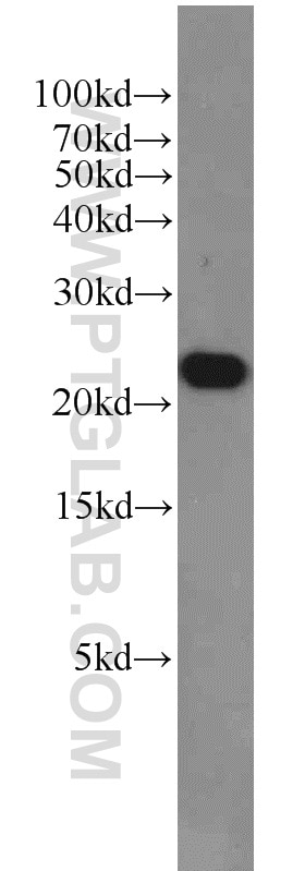
 at dilution of 1:20000 incubated at room temperature for 1.5 hours.")
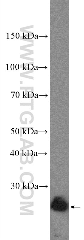
 were subjected to SDS PAGE followed by western blot with <a class='green' href='/productredirect?CatalogNo=66264-1-Ig' target='_blank'>66264-1-Ig</a> (Cytochrome c antibody) at dilution of 1:5000 incubated at room temperature for 1.5 hours.")
 at dilution of 1:8000 incubated at room temperature for 1.5 hours.")
 at dilution of 1:16000 incubated at room temperature for 1.5 hours.")
 at dilution of 1:40000 incubated at room temperature for 1.5 hours.")
 at dilution of 1:10000 incubated at room temperature for 1.5 hours.")
 with sh-Control and sh-Cytochrome c transfected <a class='green' href='/productredirect?CatalogNo=HEK-293' target='_blank'>HEK-293</a> cells.")
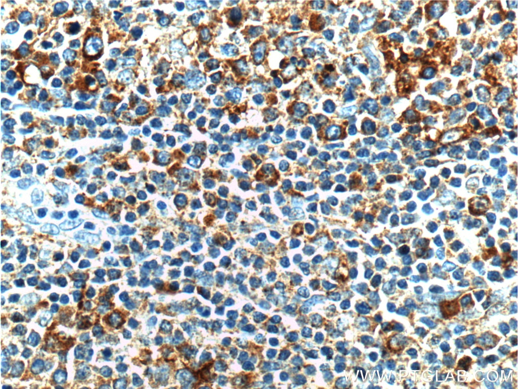
 at dilution of 1:2000 (under 10x lens). Heat mediated antigen retrieval with Tris-EDTA buffer (pH 9.0).")
 at dilution of 1:5000 (under 10x lens). Heat mediated antigen retrieval with Tris-EDTA buffer (pH 9.0).")
 at dilution of 1:5000 (under 40x lens). Heat mediated antigen retrieval with Tris-EDTA buffer (pH 9.0).")
 at dilution of 1:5000 (under 10x lens). Heat mediated antigen retrieval with Tris-EDTA buffer (pH 9.0).")
 at dilution of 1:5000 (under 40x lens). Heat mediated antigen retrieval with Tris-EDTA buffer (pH 9.0).")
 at dilution of 1:2000 (under 40x lens). Heat mediated antigen retrieval with Tris-EDTA buffer (pH 9.0).")
 fixed <a class='green' href='/productredirect?CatalogNo=HepG2' target='_blank'>HepG2</a> cells using <a class='green' href='/productredirect?CatalogNo=66264-1-Ig' target='_blank'>66264-1-Ig</a> (Cytochrome c antibody) at dilution of 1:100 and <a class='green' href='/productredirect?CatalogNo=CoraLite48' target='_blank'>CoraLite48</a>8-Conjugated AffiniPure Goat Anti-Mouse IgG(H+L). Cells were co-stained with phalloidin in red.")
 fixed <a class='green' href='/productredirect?CatalogNo=HepG2' target='_blank'>HepG2</a> cells using <a class='green' href='/productredirect?CatalogNo=66264-1-Ig' target='_blank'>66264-1-Ig</a>(Cytochrome c antibody) at dilution of 1:100 and Alexa Fluor 488-conjugated AffiniPure Goat Anti-Mouse IgG(H+L).")
 and CoraLite®488-Conjugated AffiniPure Goat Anti-Mouse IgG(H+L) at dilution 1:1000 (green), and 0.2 ug Mouse <a class='green' href='/productredirect?CatalogNo=IgG2' target='_blank'>IgG2</a>a Isotype Control (<a class='green' href='/productredirect?CatalogNo=66360-2-Ig' target='_blank'>66360-2-Ig</a>, Clone: <a class='green' href='/productredirect?CatalogNo=K11' target='_blank'>K11</a><a class='green' href='/productredirect?CatalogNo=A1' target='_blank'>A1</a><a class='green' href='/productredirect?CatalogNo=B2' target='_blank'>B2</a><a class='green' href='/productredirect?CatalogNo=A2' target='_blank'>A2</a>) (black). Cells were fixed with 4% PFA and permeabilized with 0.1% <a class='green' href='/productredirect?CatalogNo=TritonX-100' target='_blank'>TritonX-100</a>.")
Tested Applications
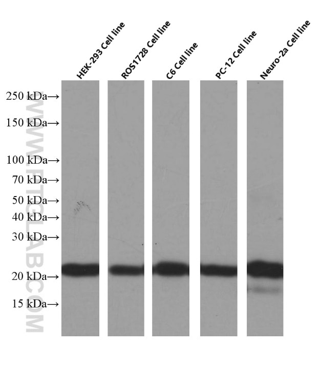
| Positive WB detected in | HeLa cells, HEK-293 cells, human heart tissue, human skeletal muscle tissue, rat skeletal muscle tissue, mouse skeletal muscle tissue, MCF-7 cells, Jurkat cells, HSC-T6 cells, ROS1728 cells, RAW 264.7 cells |
| Positive IHC detected in | human liver cancer tissue, human breast cancer tissue Note: suggested antigen retrieval with TE buffer pH 9.0; (*) Alternatively, antigen retrieval may be performed with citrate buffer pH 6.0 |
| Positive IF detected in | HepG2 cells |
| Positive FC detected in | HepG2 cells |
Recommended dilution
| Application | Dilution |
|---|---|
| Western Blot (WB) | WB : 1:5000-1:50000 |
| Immunohistochemistry (IHC) | IHC : 1:1000-1:5000 |
| Immunofluorescence (IF) | IF : 1:20-1:200 |
| Flow Cytometry (FC) | FC : 0.20 ug per 10^6 cells in a 100 µl suspension |
| Sample-dependent, check data in validation data gallery | |
Published Applications
| WB | See 86 publications below |
| IF | See 12 publications below |
Product Information
66264-1-Ig targets Cytochrome c in WB, IHC, IF, FC, ELISA applications and shows reactivity with human, mouse, rat samples.
| Tested Reactivity | human, mouse, rat |
| Cited Reactivity | human, rat, mouse, canine |
| Host / Isotype | Mouse / IgG2a |
| Class | Monoclonal |
| Type | Antibody |
| Immunogen | Cytochrome c fusion protein Ag24349 相同性解析による交差性が予測される生物種 |
| Full Name | cytochrome c, somatic |
| Calculated molecular weight | 12 kDa |
| Observed molecular weight | 12-15 kDa |
| GenBank accession number | BC009578 |
| Gene symbol | CYCS |
| Gene ID (NCBI) | 54205 |
| RRID | AB_2716798 |
| Conjugate | Unconjugated |
| Form | Liquid |
| Purification Method | Protein A purification |
| Storage Buffer | PBS with 0.02% sodium azide and 50% glycerol pH 7.3. |
| Storage Conditions | Store at -20°C. Stable for one year after shipment. Aliquoting is unnecessary for -20oC storage. |
Background Information
Cytochrome c is a 12-15 kDa electron transporting protein located in the inner mitochondrial membrane. Upon apoptotic stimulation, cytochrome c can be released from mitochondria into cytoplasm, resulting in caspase-3 activation and apoptosis. Measurement of cytochrome c release from the mitochondria is useful for detection of the onset of apoptosis in cells. In addition, cytochrome c can also leave cells and be detectable in extra-cellular medium of apoptotic cells and serum of cancer patients. The level of serum cytochrome c may serve as a prognostic maker during cancer therapy.
Protocols
| Product Specific Protocols | |
|---|---|
| WB protocol for Cytochrome c antibody 66264-1-Ig | Download protocol |
| IHC protocol for Cytochrome c antibody 66264-1-Ig | Download protocol |
| IF protocol for Cytochrome c antibody 66264-1-Ig | Download protocol |
| Standard Protocols | |
|---|---|
| Click here to view our Standard Protocols |
Publications
| Species | Application | Title |
|---|---|---|
Hepatology Hepatic stimulator substance resists hepatic ischemia/reperfusion injury by regulating Drp1 translocation and activation. | ||
Nat Commun Endonuclease G promotes autophagy by suppressing mTOR signaling and activating the DNA damage response. | ||
Redox Biol Mecheliolide elicits ROS-mediated ERS driven immunogenic cell death in hepatocellular carcinoma. | ||
J. Pineal Res. Human transporters, PEPT1/2, facilitate melatonin transportation into mitochondria of cancer cells: an implication of the therapeutic potential. | ||
Sci China Life Sci Melatonin decreases GSDME mediated mesothelial cell pyroptosis and prevents peritoneal fibrosis and ultrafiltration failure | ||
Br J Pharmacol Substitution of SERCA2 Cys674 accelerates aortic aneurysm by inducing endoplasmic reticulum stress and promoting cell apoptosis. |
Reviews
The reviews below have been submitted by verified Proteintech customers who received an incentive forproviding their feedback.
FH Megha (Verified Customer) (07-10-2018) |
|